📞Customer Service: +86 13248368268 📧servicecenter@suzhoufrank.com one year replacement and warranty!

3D Medical Imaging and Diagnostics: Benefits, Uses, and Limits
What can 3D diagnostic imaging do for your facility? Explore benefits like depth perception and precision , plus limitations hospitals should consider first.
MEDICAL IMAGING
Dr Qi Rui
12/26/20257 min read
Three-dimensional visualization has changed how clinicians see inside the human body. Traditional imaging flattens complex anatomy onto two-dimensional screens , forcing physicians to mentally reconstruct depth from flat images. 3D medical imaging eliminates this cognitive step by presenting anatomy with natural spatial relationships intact.
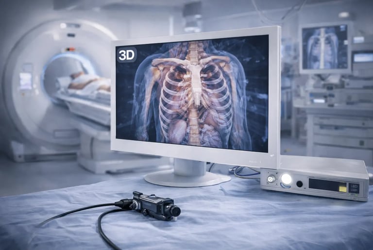
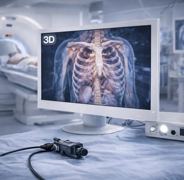
The technology spans multiple applications. Diagnostic radiology uses 3D reconstructions to visualize tumors , vascular structures and complex anatomical relationships. Surgical teams use real-time 3D visualization during minimally invasive procedures. Endoscopy increasingly incorporates stereoscopic imaging for improved navigation and tissue assessment.
But 3D imaging isn't universally superior to 2D. It adds cost , complexity and in some cases offers minimal clinical advantage over conventional approaches. Understanding where 3D diagnostic technology provides genuine benefit , and where it doesn't , helps facilities make informed investment decisions.
How 3D Medical Imaging Works
The fundamental principle behind 3D imaging is straightforward: capture visual information from two slightly different perspectives , then present those perspectives separately to each eye. The brain fuses these two views into a single image with perceived depth , just as it does with natural binocular vision.
Stereoscopic Capture
In surgical and endoscopic applications , 3D capture typically uses dual cameras or a single camera with specialized optics that split the image into left and right channels. The two perspectives are offset by roughly the same distance as human eyes (approximately 65mm) , creating natural parallax.
The captured stereo pair travels through the imaging chain as two synchronized video streams. Processing must maintain precise synchronization. Any timing mismatch between left and right channels causes visual discomfort and defeats the purpose of stereoscopic presentation.
3D Display Technologies
Several technologies present stereoscopic images to viewers. Passive 3D systems use polarized glasses and specially designed monitors that display left and right images with different polarization. Active 3D systems use shutter glasses synchronized with displays that alternate rapidly between left and right frames.
Autostereoscopic displays eliminate glasses entirely by using lenticular lenses or parallax barriers to direct different images to each eye based on viewing position. These systems offer convenience but typically restrict optimal viewing angles and positions.
Each approach involves tradeoffs between image quality , viewer comfort , cost and practical constraints like the need for special eyewear in sterile surgical environments.
Volumetric 3D Reconstruction
Beyond real-time stereoscopic visualization , 3D medical imaging includes volumetric reconstruction from cross-sectional data. CT and MRI scanners acquire hundreds of two-dimensional slices that software assembles into three-dimensional models.
These reconstructions allow clinicians to rotate , slice and examine anatomy from any angle. Surgeons use them for preoperative planning , visualizing the spatial relationships between tumors and critical structures before making incisions. Understanding advanced medical imaging technology in broader context helps appreciate how 3D reconstruction fits within the visualization landscape.
Benefits of 3D Diagnostic Imaging
The advantages of 3D medical imaging stem primarily from restored depth perception and improved spatial understanding. These benefits translate to clinical value in specific ways.
Enhanced Depth Perception
The most immediate benefit is accurate depth perception. In 2D visualization , surgeons estimate depth from indirect cues: relative size , shadows , motion parallax and tissue deformation. These cues require interpretation and can mislead.
3D imaging provides direct depth information. Surgeons see how far instruments are from tissue surfaces without estimation. They perceive the spatial relationships between structures accurately. This reduces the cognitive load of mentally translating flat images into three-dimensional reality.
Research published in The Lancet demonstrated that 3D laparoscopy improved surgical performance compared to 2D in controlled studies. Surgeons completed tasks faster and with fewer errors when using stereoscopic visualization. The benefit was particularly notable for surgeons with less experience , suggesting 3D imaging helps flatten the learning curve for complex procedures.
Improved Surgical Precision
Better depth perception translates to more precise instrument control. Suturing , dissection and tissue manipulation all benefit from accurate spatial awareness. Surgeons can place sutures more precisely , identify tissue planes more easily and navigate around critical structures with greater confidence.
In microsurgery , where operative fields are measured in millimeters , depth perception becomes even more critical. Microvascular anastomosis requires placing sutures with submillimeter precision. 3D microscopy systems provide the spatial information needed for this delicate work.
Faster Skill Acquisition
Training surgeons in minimally invasive techniques traditionally required extensive practice to develop the ability to work effectively with 2D visualization. Trainees had to learn to interpret depth from indirect cues , a skill that takes time to develop.
3D imaging reduces this learning curve. Trainees can apply their natural depth perception rather than learning to work without it. Studies have shown reduced training time and faster proficiency development when surgeons learn procedures using 3D visualization.
Better Team Communication
When the entire surgical team sees the same 3D view , communication improves. Assistants understand what the primary surgeon sees. Nurses can anticipate instrument needs based on visible spatial relationships. The shared visual reference reduces misunderstandings and improves coordination.
Traditional microscopy provided 3D views only to the surgeon looking through the eyepieces. Digital 3D microscopes display stereoscopic images on monitors visible to the entire team , democratizing access to depth information.
Clinical Applications of 3D Medical Imaging
3D diagnostic and surgical imaging has found application across multiple specialties , though adoption varies based on clinical benefit and practical considerations.
Laparoscopic and Robotic Surgery
Minimally invasive abdominal surgery was an early adopter of 3D visualization. Laparoscopic procedures performed through small incisions rely entirely on video imaging. Adding depth perception to this video feed addresses one of the fundamental limitations of the technique.
Robotic surgical systems like the da Vinci platform have used 3D visualization from their inception. The console where surgeons control robotic instruments presents stereoscopic views from dual cameras on the robotic endoscope. Surgeons consistently cite 3D visualization as a key advantage of robotic platforms.
According to the American College of Surgeons , robotic surgery continues expanding across general surgery applications , driven partly by visualization advantages including 3D imaging and magnification.
Endoscopy
3D endoscopy has developed more recently. Traditional endoscopes used single cameras providing 2D views. Newer systems incorporate stereoscopic capture for procedures where depth perception offers clinical advantage.
Endoscopic submucosal dissection (ESD) , a technique for removing early GI cancers without surgery , benefits from 3D visualization. The procedure requires precise depth control to dissect within the submucosal layer without perforating the bowel wall. Stereoscopic imaging helps endoscopists maintain correct tissue plane.
Bronchoscopy and thoracoscopy applications similarly benefit when navigating complex three-dimensional anatomy or performing interventions requiring precise depth control.
Neurosurgery
Neurosurgical microscopes have long provided stereoscopic visualization through optical binoculars. Digital 3D microscopes extend this capability to the entire operative team while adding recording and integration features.
Beyond real-time operative visualization , 3D reconstruction from preoperative MRI and CT plays a crucial role in neurosurgical planning. Surgeons visualize the spatial relationships between tumors , vessels and eloquent brain regions before operating. Image-guided navigation systems use these 3D models to provide intraoperative guidance.
Diagnostic Radiology
Radiologists increasingly use 3D reconstructions for diagnostic interpretation. Complex vascular anatomy , fracture patterns and tumor relationships to surrounding structures often become clearer when viewed in three dimensions.
3D diagnostic imaging proves particularly valuable for surgical planning consultations. When radiologists confer with surgeons about operative approach , 3D models provide a shared visual reference that communicates spatial relationships more effectively than 2D slices.
Cardiac imaging relies heavily on 3D reconstruction to visualize heart chambers , valves and coronary arteries. The dynamic , three-dimensional nature of cardiac anatomy makes 3D visualization particularly valuable.
Limitations and Considerations
Despite genuine benefits , 3D medical imaging has limitations that facilities should understand before investing.
Cost Premium
3D imaging systems cost more than 2D equivalents. Stereoscopic cameras require dual sensors and more complex optics. 3D displays add expense regardless of the technology used. The overall system cost increase typically ranges from 20% to 50% or more depending on the platform.
This premium must be justified by clinical benefit. For procedures where depth perception significantly affects outcomes or efficiency , the investment may pay for itself. For procedures where 2D visualization suffices , the added cost provides little return.
Visual Fatigue
Extended viewing of stereoscopic displays can cause visual fatigue , headaches and discomfort in some users. The phenomenon varies among individuals. Some surgeons experience no issues with prolonged 3D use. Others find fatigue limits how long they can work effectively with stereoscopic visualization.
Display quality affects fatigue levels. High-quality 3D systems with properly calibrated stereo geometry and accurate color matching between channels cause less strain than lower-quality implementations. Crosstalk (leakage of left-eye images into the right eye and vice versa) particularly contributes to discomfort.
Learning Curve
While 3D imaging reduces the learning curve for spatial tasks , it introduces its own adaptation period. Surgeons accustomed to 2D visualization have developed compensatory skills over years of practice. Switching to 3D requires recalibration.
Some experienced surgeons prefer 2D systems they've mastered over 3D alternatives that feel unfamiliar. This preference should be respected. Forcing 3D technology on surgeons who work effectively with 2D may not improve outcomes.
Technical Complexity
3D systems have more components that can fail. Synchronization between channels must be maintained. Display calibration must preserve stereo geometry. Glasses or headsets must be managed , cleaned and maintained.
This added complexity increases maintenance requirements and potential failure points. Facilities adopting 3D technology need technical support capabilities to keep systems functioning properly.
Not Always Superior
Perhaps the most important limitation is that 3D imaging doesn't universally outperform 2D. For many diagnostic applications , 2D imaging provides all necessary information. For some surgical tasks , depth perception matters less than other factors.
Evidence for clinical outcome improvement with 3D imaging remains mixed depending on application. Performance on simulated tasks improves consistently. Translation to patient outcomes is less clearly established for all procedure types.
According to research in PMC , while 3D laparoscopy demonstrates performance advantages in experimental settings , the magnitude of clinical benefit varies by procedure complexity and surgeon experience. The technology offers the most advantage in technically demanding procedures with significant depth-related challenges.
Making the Decision
Facilities considering 3D medical imaging investments should evaluate several factors.
Procedure Mix
What procedures will use the technology? Complex laparoscopic surgery , robotic procedures , microsurgery and certain endoscopic interventions benefit most from 3D visualization. Facilities with high volumes of these procedures have stronger justification for investment.
Surgeon Preferences
Will the surgeons who'll use the equipment embrace 3D technology? Early adopters eager to try new visualization tools will maximize the investment. Surgeons satisfied with current 2D systems may not utilize 3D capabilities even when available.
Training Programs
Facilities with surgical training programs may find particular value in 3D imaging. Reducing the learning curve for trainees accelerates skill development and potentially improves patient safety during the training period.
Budget Constraints
The cost premium for 3D capability must fit within equipment budgets. If choosing between a high-quality 2D system and a marginal 3D system at the same price point , the 2D option may serve patients better.
The Future of 3D Diagnostic Imaging
3D medical imaging continues evolving. Emerging developments include better autostereoscopic displays that eliminate glasses without sacrificing quality , integration with augmented reality for 3D overlay of imaging data onto surgical fields , and AI-enhanced 3D reconstruction that generates detailed models from limited input data.
As technology improves and costs decrease , 3D imaging will likely become standard for more applications. Facilities investing in current 3D platforms should consider upgrade paths and compatibility with emerging technologies.
The trajectory is clear: three-dimensional visualization addresses a fundamental limitation of traditional medical imaging. Where that limitation matters clinically , 3D technology provides genuine value. Making informed decisions about when and where to deploy 3D imaging ensures facilities invest wisely in visualization capabilities that improve patient care.
© 2025. All rights reserved.
About Us
Introduction
Development
Cooperation
Service
Main Products
Medical Grade Monitor
No 15, Jinyang road KunshanSuzhou, Jiangsu, China