📞Customer Service: +86 13248368268 📧servicecenter@suzhoufrank.com one year replacement and warranty!

Advanced Medical Imaging Technology in Modern Surgery
How advanced medical imaging shapes modern surgery. From 4K cameras to surgical displays and 3D microscopes , understand the technology behind better visualization.
MEDICAL IMAGING
Dr Qi Rui
12/25/202512 min read
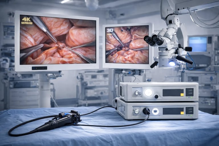
Advanced medical imaging has fundamentally changed how surgery is performed. What was once guided primarily by direct vision and tactile feedback now relies heavily on sophisticated visualization systems that reveal anatomy in ways the human eye alone cannot perceive. From high-resolution cameras to precision displays and optical microscopes , medical imaging technology forms the visual backbone of modern surgical practice.
For healthcare facilities evaluating their surgical capabilities , understanding this technology isn't optional. It's essential. The quality of visualization directly impacts surgical precision , procedural safety and patient outcomes. Surgeons operating with substandard imaging face unnecessary challenges. Those equipped with advanced systems can see more , do more and achieve better results.
This guide examines the core concepts behind advanced medical imaging in surgical settings. It covers the fundamental components , explains why specifications like resolution and latency matter , and provides the foundation for informed equipment decisions.
Why Visualization Quality Matters in Surgery
The importance of visualization in surgery cannot be overstated. Surgeons make split-second decisions based on what they see. When imaging systems fail to deliver clear , accurate , real-time visual information , the consequences can be significant.
Consider minimally invasive surgery. The surgeon isn't looking directly at the operative field. Instead , they're watching a monitor displaying video from an endoscopic camera. Every aspect of that imaging chain affects what the surgeon perceives: the camera's resolution , the display's color accuracy , the system's processing speed , the monitor's brightness in a brightly lit OR. Any weakness in this chain compromises the surgeon's ability to distinguish tissue types , identify anatomical structures and detect subtle abnormalities.
The World Health Organization recognizes medical imaging as playing a central role in modern medicine , supporting both diagnosis and treatment of disease. This recognition extends to surgical applications where imaging guides interventions in real time. Diagnostic medical imaging and clinical insight work together to inform treatment decisions before , during and after surgical procedures.
Beyond minimally invasive techniques , advanced imaging supports open surgery through microscopes that reveal cellular-level detail , display systems that present radiological images alongside live video , and integration platforms that synthesize multiple data sources into coherent visual information. The trend across all surgical specialties points toward greater reliance on technology-mediated visualization.
Core Components of Surgical Imaging Systems
Every surgical imaging system comprises several interconnected components. Understanding each element helps facilities make informed purchasing decisions and optimize existing equipment.
Image Capture Devices
The imaging chain begins with capture. In endoscopic and laparoscopic surgery , this means cameras mounted on flexible or rigid scopes. In microsurgery , optical microscopes provide magnified views. In image-guided procedures , real-time fluoroscopy or ultrasound generates the visual feed.
Camera technology has advanced dramatically. Modern surgical cameras use either CCD (charge-coupled device) or CMOS (complementary metal-oxide semiconductor) sensors. Both convert light into electronic signals , but they differ in power consumption , heat generation and image characteristics. CMOS sensors have gained ground recently due to improvements in image quality and advantages in miniaturization.
The quality of the captured image depends on sensor resolution , light sensitivity , dynamic range and color reproduction. Higher-end cameras capture more detail , perform better in varying light conditions and reproduce colors more accurately. These specifications translate directly to what surgeons see on their displays.
Light Sources
Adequate illumination is fundamental. Internal body cavities are dark. Without sufficient light , even the best camera produces unusable images.
Traditional light sources used halogen or xenon bulbs. These provided acceptable illumination but generated significant heat , consumed substantial power and required regular bulb replacement. Modern LED light sources have largely replaced older technologies , offering longer lifespan (often exceeding 30,000 hours) , lower heat output , instant-on capability and more consistent color temperature over time.
Light intensity must be sufficient to illuminate deep anatomical structures while avoiding overexposure of proximal tissues. Adjustable output allows optimization for specific procedures and patient anatomies.
Medical Displays
The display translates captured images into visual information for the surgical team. Medical-grade monitors differ substantially from consumer displays in ways that matter clinically.
Surgical displays must perform in challenging environments: bright ambient lighting from surgical lamps , viewing by multiple team members from various angles , continuous operation for extended procedures. They require high brightness (often exceeding 500 cd/m²) , wide viewing angles , accurate color reproduction and resistance to glare.
Resolution matters significantly. Understanding how to choose a medical display for surgery involves evaluating resolution alongside other specifications. A display that can't resolve the detail captured by a 4K camera wastes the camera's capability. Matching display resolution to camera output optimizes the imaging investment.
What is a medical-grade monitor exactly? Beyond resolution and brightness , medical-grade certification ensures devices meet safety standards for use near patients , withstand hospital cleaning protocols and maintain consistent performance over time.
Signal Processing and Transmission
Between camera and display , signals must be processed and transmitted. This involves video processors , cables , switches and sometimes network infrastructure.
Processing includes color correction , image enhancement , noise reduction and format conversion. Modern processors perform these functions in real time without introducing perceptible delay. Advanced features may include image stabilization , contrast optimization and integration of multiple video sources.
Transmission standards affect both quality and latency. SDI (Serial Digital Interface) connections , particularly 12G-SDI for 4K content , provide reliable high-bandwidth transmission over coaxial cables. HDMI and DisplayPort connections offer alternatives with their own advantages and limitations.
Resolution: From Standard Definition to 4K and Beyond
Resolution describes how much detail an imaging system can capture and display. Higher resolution means more pixels , which translates to finer detail visibility.
Standard Definition (SD)
Older endoscopic systems operated at approximately 480 lines of resolution. While adequate for basic visualization , SD systems struggle to reveal subtle findings. Fine vascular patterns , early mucosal changes and small lesions may escape detection at SD resolution. Many facilities worldwide still operate SD equipment , representing aging infrastructure in need of modernization.
High Definition (HD)
HD systems deliver 720p (1280 × 720 pixels) or 1080p (1920 × 1080 pixels) resolution. The improvement from SD to HD is substantial and clinically meaningful. Studies have demonstrated that HD endoscopy detects more adenomas during colonoscopy compared to SD equipment. For surgical applications , HD provides significantly better tissue differentiation and anatomical detail.
HD remains the current baseline for acceptable surgical visualization. Facilities operating below HD resolution should prioritize upgrades.
4K Ultra High Definition
4K systems offer approximately 3840 × 2160 pixels , four times the resolution of 1080p HD. The increase in detail visibility is considerable. Fine structures , subtle color variations and minute anatomical features become visible with 4K that HD might miss.
According to research published in PMC , advances in medical imaging technology including higher-resolution systems have enhanced visualization and contributed to improved surgical outcomes. The combination of AI-driven imaging with real-time high-resolution visualization provides surgeons with augmented decision support during procedures.
For procedures requiring maximum visual information , such as complex oncological resections or delicate neurosurgical work , 4K imaging offers meaningful advantages. Selecting the best endoscopy and surgical monitors means evaluating 4K capability alongside other specifications.
3D Visualization
Resolution describes two-dimensional detail. 3D imaging adds depth perception , fundamentally changing how surgeons perceive the operative field.
Traditional 2D displays flatten three-dimensional anatomy onto a two-dimensional screen. Surgeons mentally reconstruct depth from visual cues like shadows , relative size and motion parallax. This works but imposes cognitive load.
3D systems capture stereoscopic images using dual cameras or specialized optics and present them on displays that recreate depth perception. Surgeons see the operative field with natural spatial relationships , improving hand-eye coordination and reducing the mental effort required to navigate three-dimensional anatomy.
The benefits and limitations of 3D medical imaging and diagnostics continue to be studied , but clinical adoption is growing , particularly in robotic surgery and complex laparoscopic procedures.
Microscopy in Modern Surgery
Surgical microscopes represent another critical category of advanced medical imaging technology. Where endoscopic cameras capture video from within body cavities , microscopes provide magnified views of exposed surgical fields.
Optical Principles
Surgical microscopes use precision optics to magnify anatomy from approximately 4× to 40× or higher. Variable magnification allows surgeons to zoom in for fine work and pull back for orientation. High-quality optics deliver sharp , distortion-free images across the entire field of view.
Illumination integrates directly with the optical path , ensuring the magnified field receives adequate light. Modern microscopes use LED or xenon illumination with adjustable intensity.
Applications
Neurosurgery relies heavily on surgical microscopes for procedures involving delicate neural structures. Ophthalmology uses specialized microscopes for intraocular surgery. ENT surgery employs microscopes for middle ear procedures. Plastic and reconstructive surgery uses microscopy for microvascular anastomosis.
Understanding how to choose the right clinical microscopes requires matching optical specifications , illumination capabilities and ergonomic features to the specific surgical applications a facility performs.
3D Microscopes
Just as endoscopic systems have evolved to include 3D visualization , surgical microscopes now offer stereoscopic options. Traditional binocular microscopes provide natural stereo vision through separate eyepieces for each eye. Digital 3D microscopes capture stereo images and display them on 3D monitors , allowing the entire team to share the surgeon's view.
Medical 3D microscopes matter most when depth perception significantly impacts surgical precision , such as in microvascular surgery where suture placement occurs at the millimeter scale.
Color Accuracy: Why It Matters Clinically
Resolution captures detail. Color accuracy ensures that detail is represented truthfully. In surgical imaging , color carries diagnostic information that can be as important as structural detail.
Tissue differentiation often depends on subtle color variations. Healthy tissue versus ischemic tissue. Arterial versus venous blood. Normal mucosa versus early neoplastic change. Inflamed tissue versus healthy tissue. These distinctions frequently manifest as color differences before structural changes become apparent.
When imaging systems distort color , they compromise the surgeon's ability to make these distinctions. A display that shifts reds toward orange may mask signs of adequate perfusion. A camera that oversaturates colors may make normal tissue appear abnormal. Inaccurate color reproduction introduces uncertainty into visual assessment.
Color Standards in Medical Imaging
Medical displays are calibrated to specific color standards. The most common include sRGB for general clinical use and ITU-R BT.709 for video applications. Some specialized applications use wider color gamuts to capture more of the visible spectrum.
DICOM (Digital Imaging and Communications in Medicine) calibration applies primarily to grayscale diagnostic imaging but establishes the principle that medical displays must render information consistently and accurately. Surgical displays follow analogous principles for color accuracy.
Calibration and Maintenance
Color accuracy isn't permanent. Displays drift over time as backlights age and components degrade. Regular calibration ensures consistent performance throughout a display's service life.
High-end surgical monitors include built-in calibration sensors that automatically maintain color accuracy. Less sophisticated displays require periodic manual calibration using external colorimeters. Facilities should establish calibration protocols and schedules as part of their equipment maintenance programs.
The Radiological Society of North America emphasizes the importance of proper display calibration and quality assurance in medical imaging environments. While their guidance focuses primarily on diagnostic radiology , the principles apply equally to surgical visualization where accurate image representation affects clinical decisions.
Latency: The Invisible Performance Factor
Latency refers to the delay between an event occurring and its appearance on screen. In surgical imaging , latency measures the time from when the camera captures an image to when the surgeon sees it on the display.
Why Latency Matters
Even small delays affect surgical performance. Hand-eye coordination depends on visual feedback occurring in real time. When surgeons move an instrument and the visual confirmation arrives late , the mismatch disrupts fine motor control.
Studies of laparoscopic performance demonstrate that latency above 100-200 milliseconds measurably degrades surgical precision. At higher latencies , surgeons must slow their movements to compensate for delayed feedback , extending procedure times and increasing cognitive load.
Sources of Latency
Latency accumulates throughout the imaging chain. Camera sensor readout introduces delay. Video processing adds more. Signal transmission contributes depending on the medium and distance. Display processing and pixel response time add final increments.
Each component contributes milliseconds. Combined , these can push total system latency into ranges that affect performance. Understanding where latency originates helps facilities specify and configure systems for optimal real-time performance.
Minimizing Latency
Low-latency surgical imaging requires attention at every stage. Cameras with fast sensor readout and minimal onboard processing reduce capture delay. Direct SDI connections avoid the compression delays associated with some network-based video transmission. Displays with fast pixel response times and minimal input processing complete the chain.
Modern surgical imaging systems designed for real-time applications typically achieve end-to-end latency below 100 milliseconds. Some high-performance systems achieve latency below 50 milliseconds , effectively imperceptible to users.
Integration: Bringing Multiple Sources Together
Modern surgical environments generate visual information from multiple sources simultaneously. Endoscopic cameras provide live operative views. Fluoroscopy or ultrasound may offer real-time imaging guidance. Preoperative CT or MRI scans provide anatomical reference. Patient monitoring systems display vital signs. PACS workstations show relevant prior studies.
Surgeons need access to all this information without constantly shifting attention between separate displays scattered around the room. Integration systems consolidate multiple sources into coherent visual presentations.
Picture-in-Picture and Multi-View Displays
Basic integration involves displaying multiple video sources on a single monitor. Picture-in-picture places a smaller secondary image over the primary view. Picture-by-picture divides the screen into zones showing different sources simultaneously.
Large-format displays (55 inches and above) make multi-view configurations practical by providing sufficient screen real estate. A surgeon might view the primary endoscopic feed in the center , fluoroscopy in one corner , vital signs in another and a reference CT scan in a third zone.
Surgical Integration Platforms
Advanced integration goes beyond simple video switching. Surgical integration platforms route video from any source to any display in the OR. They record procedures for documentation and training. They enable communication with remote experts. They interface with hospital information systems to pull relevant patient data.
These platforms transform the OR into a connected environment where information flows to where it's needed. Medical imaging analysis becomes more effective when relevant comparison images are immediately accessible during procedures.
Augmented Reality Integration
Emerging technologies project imaging data directly into the surgeon's field of view. Augmented reality (AR) systems overlay preoperative imaging onto the live surgical field , showing the location of structures beneath the visible surface.
AR navigation systems register patient anatomy to preoperative scans , then display relevant information in spatial context. A surgeon might see the planned resection margin projected onto the tissue surface or the location of a tumor relative to visible landmarks.
According to the National Institutes of Health , image-guided therapy represents an emerging field with close relationships to interventional radiology , minimally invasive surgery , computer-assisted visualization and robot-assisted surgery. The integration of real-time imaging with preoperative data continues advancing surgical precision.
Innovative Trends Shaping the Future
Medical imaging technology continues evolving rapidly. Facilities planning equipment investments should understand emerging trends that may affect future capabilities and upgrade paths.
Artificial Intelligence
AI is transforming medical imaging from a passive visualization tool into an active diagnostic assistant. Machine learning algorithms analyze images in real time , highlighting regions of interest , measuring structures automatically and flagging potential abnormalities.
In surgical applications , AI can provide real-time tissue characterization , distinguishing tumor from normal tissue based on visual patterns. AI-powered systems can track instruments , measure distances and provide navigation guidance. As these capabilities mature , they'll become standard features in surgical imaging platforms.
8K Resolution
While 4K adoption continues expanding , 8K systems (7680 × 4320 pixels) are entering the market. The jump from 4K to 8K quadruples pixel count again , potentially revealing even finer anatomical detail.
Whether 8K provides clinically meaningful improvement over 4K for most surgical applications remains to be established. Display size , viewing distance and the limits of optical systems all affect whether additional pixels translate to visible benefit. However , facilities investing in infrastructure should consider future compatibility with higher-resolution systems.
Hyperspectral and Multispectral Imaging
Standard surgical cameras capture visible light in three color channels (red , green , blue). Hyperspectral imaging captures dozens or hundreds of narrow spectral bands , revealing tissue properties invisible to conventional cameras.
Hyperspectral analysis can assess tissue oxygenation , identify tissue types and potentially detect malignancy based on spectral signatures. While still largely experimental , this technology represents a direction where surgical imaging may evolve.
Robotic Integration
Robotic surgical systems rely entirely on imaging for surgeon feedback. The console where surgeons control robotic instruments presents stereoscopic video from cameras mounted on the robotic arms. Image quality directly affects the surgeon's ability to operate effectively.
As robotic surgery expands into more procedures and specialties , the integration between imaging systems and robotic platforms will deepen. Innovative medical imaging trends increasingly intersect with robotic surgery development.
Selecting Equipment for Your Facility
Choosing advanced medical imaging technology requires balancing clinical needs , budget constraints and future considerations. Several principles guide effective equipment selection.
Match Technology to Clinical Applications
Different procedures have different visualization requirements. A facility performing primarily screening colonoscopies has different needs than one specializing in complex hepatobiliary surgery. Understanding the specific visual demands of your procedure mix guides appropriate specification.
Resolution , 3D capability , color accuracy requirements and integration needs all vary by application. Specifying beyond actual clinical requirements wastes resources. Specifying below requirements compromises patient care.
Consider the Complete System
Imaging quality depends on the entire chain from camera to display. Upgrading one component while neglecting others may yield disappointing results. A 4K camera feeding an HD display provides HD images. A high-quality display receiving compressed video can't recover lost information.
Evaluate systems holistically. Ensure cameras , processors , transmission infrastructure and displays are all capable of supporting the image quality you're targeting.
Plan for Integration
Standalone imaging equipment increasingly gives way to integrated systems. Consider how new equipment will interface with existing infrastructure , recording systems , PACS and hospital networks. Proprietary systems that don't interoperate create islands of capability that can't be combined.
Open standards and common interfaces facilitate integration. Evaluate vendor commitment to interoperability when selecting equipment.
Account for Total Cost of Ownership
Purchase price represents only part of equipment cost. Maintenance contracts , consumables (like xenon bulbs for older light sources) , calibration requirements , upgrade paths and expected service life all affect total cost of ownership.
LED light sources cost more initially but eliminate bulb replacement costs. Displays with built-in calibration reduce service requirements. Equipment from established manufacturers typically offers better long-term support than budget alternatives.
Future-Proof Where Practical
Technology evolves continuously. Equipment purchased today will serve for years. Where possible , select systems that can accommodate foreseeable advances.
This might mean choosing displays that support 4K even if current cameras are HD , anticipating future camera upgrades. It might mean selecting integration platforms with expansion capacity beyond current needs. It might mean ensuring network infrastructure can handle increasing video bandwidth.
Balance future-proofing against paying for capability you may never use. Focus on likely evolution paths relevant to your clinical programs.
Partnering for Advanced Imaging Solutions
Implementing advanced medical imaging technology requires more than purchasing equipment. It requires understanding clinical workflows , integrating with existing systems , training staff and maintaining performance over time.
Suzhou Frank Medical provides comprehensive surgical imaging solutions designed for modern operating environments. Our portfolio includes HD and 4K camera systems , LED light sources , medical-grade displays and portable imaging units suitable for facilities ranging from major medical centers to ambulatory surgery centers.
Whether you're building a new surgical suite , upgrading aging equipment or expanding imaging capabilities to new locations , our team can help identify the right configuration for your clinical priorities and budget.
Contact us to discuss your advanced medical imaging needs or explore our surgical visualization products to see available options.
© 2025. All rights reserved.
About Us
Introduction
Development
Cooperation
Service
Main Products
Medical Grade Monitor
No 15, Jinyang road KunshanSuzhou, Jiangsu, China