📞Customer Service: +86 13248368268 📧servicecenter@suzhoufrank.com one year replacement and warranty!

Medical 3D Microscopes: When Depth Perception Matters
When does a 3D microscope outperform a standard medical microscope? Explore surgical applications where depth perception improves precision , safety and outcomes.
MEDICAL IMAGING
Dr Qi Rui
1/3/20267 min read
Picture a neurosurgeon navigating around a tumor millimeters from the optic nerve. Or a plastic surgeon reconnecting blood vessels thinner than a human hair. In these moments , the difference between seeing in two dimensions versus three isn't a luxury. It's the difference between success and complication.
A medical microscope has long been essential for procedures requiring magnification beyond what the naked eye can achieve. But traditional optical microscopes present a flat world. Surgeons must mentally reconstruct depth from monocular cues , shadows and experience. The emergence of the 3D microscope changes this equation , restoring natural depth perception at magnified scales where spatial relationships become critical.
This guide explores when and why depth perception matters in microscopy , how 3D technology addresses limitations of traditional systems and what facilities should consider when evaluating these instruments.
The Challenge of Working Without Depth
Humans perceive depth through binocular vision. Each eye sees a slightly different angle , and the brain fuses these images into three-dimensional understanding. This happens automatically , unconsciously , constantly.
Traditional surgical microscopes disrupt this natural process. Even binocular microscopes that provide separate eyepieces often deliver the same optical image to both eyes. The surgeon sees magnified detail but loses stereoscopic depth information precisely when it matters most.
The consequences are real. A 2019 study published by the International Society for Optics and Photonics found that surgeons operating with 2D visualization required more time for complex spatial tasks and showed increased error rates compared to those with stereoscopic visualization. The brain compensates for missing depth cues , but compensation requires cognitive effort that could otherwise focus on the procedure itself.
Experienced surgeons develop remarkable ability to work within 2D constraints. They learn to use shadows , motion parallax and anatomical knowledge to infer depth. But why impose this cognitive burden when technology can restore what flat imaging takes away?
How 3D Microscopes Restore Depth Perception
A true 3D microscope captures separate images from two slightly offset optical paths , mimicking how human eyes see the world. When these dual images reach the surgeon's visual system , the brain perceives genuine depth just as it would viewing the actual anatomy directly.
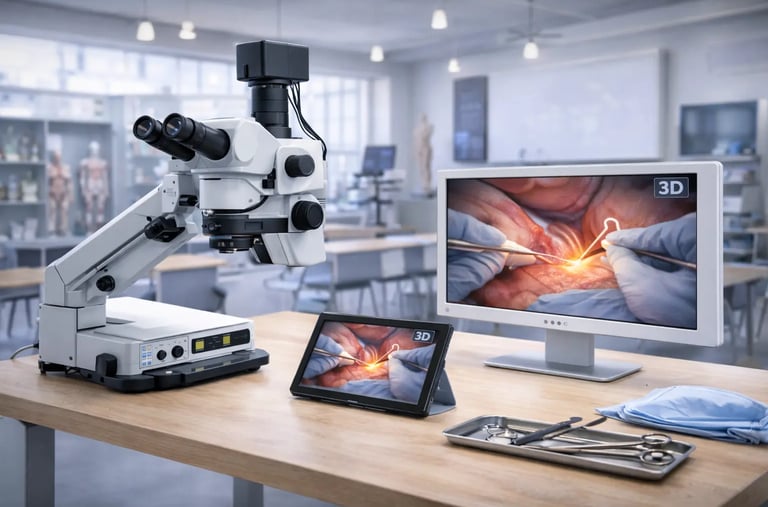
The technical approaches vary. Some systems deliver stereoscopic images through specialized eyepieces. Others project 3D visualization onto external displays. The newest generation offers naked-eye 3D viewing on integrated monitors that require no glasses or headsets.
What unites these approaches is the restoration of spatial information that 2D systems sacrifice. Tissue layers become distinguishable by depth , not just color and texture. Instrument tips can be positioned precisely relative to structures above and below. The mental effort of reconstructing three dimensions from flat images redirects toward the surgery itself.
The American Academy of Ophthalmology has documented the adoption of 3D visualization in ophthalmic surgery , noting benefits for both surgical precision and educational observation. When multiple team members can view procedures with depth perception intact , learning accelerates.
Where Depth Perception Matters Most
Not every application of a medical microscope demands 3D capability. But certain procedures benefit dramatically from restored depth perception.
Neurosurgery
Operating within the brain requires navigating complex three-dimensional anatomy where critical structures lie in different planes. Tumors may wrap around vessels. Lesions may abut eloquent cortex. A 3D microscope helps surgeons understand these spatial relationships in real time rather than reconstructing them mentally.
Skull base surgery particularly benefits. Approaching deep structures through narrow corridors demands precise understanding of where instruments are relative to surrounding anatomy. Depth perception reduces the risk of inadvertent contact with structures outside the intended surgical path.
Ophthalmic Surgery
The eye presents layered structures in a confined space. Cataract surgery , vitreoretinal procedures and corneal work all involve manipulating tissues at different depths within a small anatomical volume. Stereoscopic visualization helps surgeons understand exactly where their instruments are within this layered environment.
Microsurgery and Reconstruction
Reconnecting severed nerves or anastomosing tiny vessels requires working at scales where depth errors of fractions of a millimeter matter. Plastic surgeons and hand surgeons performing microsurgical reconstruction benefit from 3D visualization that reveals true spatial relationships between structures being joined.
ENT and Head/Neck Procedures
Otologic surgery , sinus procedures and other ENT applications involve complex anatomy with critical structures in close proximity. A 3D microscope helps surgeons navigate these intricate spaces with greater confidence in spatial positioning.
Beyond the Eyepieces: External 3D Displays
Traditional microscope eyepieces limit visualization to whoever is positioned at the oculars. Everyone else in the room , assistants , nurses , trainees , sees nothing or watches a flat 2D monitor feed.
Modern 3D microscope systems address this limitation by outputting stereoscopic video to external displays. This fundamentally changes how surgical teams interact with microscope-guided procedures.
When paired with quality 3D visualization displays , the entire team sees what the surgeon sees , depth included. Assistants can anticipate needs more effectively when they understand the three-dimensional context. Trainees learn faster when they perceive spatial relationships directly rather than trying to infer them from flat images.
Naked-eye 3D technology takes this further by eliminating the glasses that traditional 3D displays require. Suzhou Frank Medical's naked-eye 3D microscope system exemplifies this approach , providing glasses-free stereoscopic visualization that multiple observers can view simultaneously without special eyewear.
For facilities building comprehensive visualization infrastructure , understanding how microscopy integrates with broader advanced medical imaging technology systems helps ensure cohesive investment decisions.
Portable 3D Visualization for Training and Review
Surgical training traditionally required being physically present in the operating room , positioned where visualization was possible. A trainee who couldn't see couldn't learn.
3D microscope systems with recording capability change this dynamic. Captured stereoscopic footage can be reviewed later with full depth perception preserved. Complex portions of procedures can be studied repeatedly. Rare cases can become teaching resources that benefit learners who weren't present for the original surgery.
Portable display options extend this capability further. 3D tablet devices allow stereoscopic review outside the operating room. Morning conferences can include 3D case presentations. Fellows can study at home with the same depth visualization they would have at the microscope.
The educational implications are significant. Skills that previously required extensive in-person observation can now be developed through recorded stereoscopic content. This doesn't replace hands-on experience but supplements it in ways that 2D recording never could.
Ergonomic Considerations
Hours spent at microscope eyepieces take a physical toll. Neck strain , back problems and eye fatigue affect surgeons who perform microscope-dependent procedures regularly. The Congress of Neurological Surgeons has addressed ergonomic concerns in surgical practice , recognizing that equipment design affects long-term physician wellbeing.
3D microscope systems with external display options offer ergonomic alternatives to traditional eyepiece-bound postures. Surgeons can operate while viewing a 3D monitor positioned at comfortable height and distance. Heads-up surgery , as this approach is sometimes called , allows more natural posture during lengthy procedures.
The transition requires adjustment. Surgeons accustomed to eyepiece work must adapt to monitor-based visualization. But many who make the switch report reduced fatigue and find the ergonomic benefits worth the learning curve.
Naked-eye 3D displays eliminate an ergonomic burden that glasses-based 3D systems impose. Wearing 3D glasses over prescription lenses , or for extended periods , creates discomfort that naked-eye technology avoids entirely.
Image Quality and the 3D Experience
Stereoscopic visualization only delivers value when underlying image quality supports it. A 3D microscope with poor resolution or color accuracy provides depth perception of inadequate images. Both dimensions of quality matter.
Resolution determines how much detail is visible. 4K capture and display reveal fine structures that lower resolutions blur or obscure. When working at microscopic scales , this detail can be clinically significant.
Color accuracy ensures tissues appear as they actually are. Subtle color differences between tissue types , between healthy and pathological tissue , provide diagnostic information. Display systems must render these differences faithfully.
Brightness and contrast affect visibility in surgical lighting environments. Displays that wash out under OR lighting or fail to show subtle tonal differences compromise the visualization they're meant to provide.
Facilities evaluating 3D microscope systems should assess the complete imaging chain. Camera quality , processing capability , transmission fidelity and display performance all contribute to the final visual experience.
When 2D Remains Sufficient
Not every procedure requires 3D visualization. Acknowledging this helps facilities invest appropriately rather than over-specifying equipment.
Dermatologic examination , routine pathology review and many diagnostic applications work well with conventional medical microscope systems. When depth perception doesn't affect clinical decisions , 3D capability adds cost without proportional benefit.
The question to ask: does this application involve spatial relationships where depth ambiguity could affect outcomes or efficiency? If yes , 3D technology deserves consideration. If the work is fundamentally about surface examination or single-plane evaluation , traditional systems may serve equally well.
Making the Investment Decision
Facilities considering 3D microscope technology should evaluate several factors:
Clinical Applications
What procedures will use the system? Do those procedures involve depth-critical spatial relationships? Would surgeons currently performing those procedures benefit from stereoscopic visualization?
Team Visualization Needs
Is shared visualization important? Do assistants , trainees or observers need to see what the primary surgeon sees? External 3D display capability matters more when multiple viewers are involved.
Ergonomic Priorities
Are surgeons experiencing physical strain from current equipment? Would heads-up surgery options address those concerns? Ergonomic benefits may justify investment even when pure visualization needs don't.
Training Programs
Does the facility train residents or fellows? Would stereoscopic recording and playback capability enhance educational programs? Training value can factor into investment justification.
Integration Requirements
How will new microscopy equipment integrate with existing imaging infrastructure , recording systems and display networks? Compatibility with current systems affects implementation complexity.
Looking Ahead
The trajectory of medical microscope technology points toward increasingly sophisticated 3D visualization becoming standard rather than exceptional. As costs decrease and benefits become better documented , adoption will accelerate.
Facilities investing now position themselves ahead of this curve. They gain experience with 3D workflows while the technology remains differentiating. They build libraries of stereoscopic educational content. They attract surgeons who prefer working with advanced visualization.
Those who wait face no immediate penalty but may find themselves catching up rather than leading as 3D microscopy becomes expected capability for complex procedures.
Conclusion
Depth perception isn't a feature. It's how humans naturally see the world. Traditional medical microscope systems sacrifice this natural vision at precisely the moments when spatial understanding matters most. The 3D microscope restores what flat imaging takes away , giving surgeons back the depth perception their brains expect.
For procedures involving complex three-dimensional anatomy , for teams that benefit from shared stereoscopic visualization , for facilities prioritizing surgical education , 3D microscopy offers advantages that 2D systems cannot match. The question isn't whether depth perception matters. It's whether your applications are among those where it matters most.
© 2025. All rights reserved.
About Us
Introduction
Development
Cooperation
Service
Main Products
Medical Grade Monitor
No 15, Jinyang road KunshanSuzhou, Jiangsu, China