📞Customer Service: +86 13248368268 📧servicecenter@suzhoufrank.com one year replacement and warranty!

Capsule Endoscopy: How It Works
Capsule endoscopy is key for finding obscure GI bleeding & Crohn's disease in the small bowel. See preparation, what the camera records, and next steps.
ENDOSCOPY
Dr Qi Rui
12/17/20259 min read
Capsule endoscopy is a diagnostic procedure that uses a tiny wireless camera to photograph your digestive tract from the inside. You swallow a pill-sized capsule. It travels through your system taking thousands of pictures. And those images get transmitted to a recorder you wear on your belt.
Pretty simple concept. But the technology has transformed how physicians examine the small intestine.
Here's the thing. The small intestine stretches about 20 feet long. Traditional endoscopes can't reach most of it. Before capsule endoscopy came along, this entire section was essentially a blind spot. Now doctors can see the whole thing in remarkable detail. No sedation. No discomfort. No hanging around a medical facility all day.
If you want to understand endoscopy more broadly, our guide on what is endoscopy covers the basics. This article focuses specifically on the capsule version.
What Exactly Is Capsule Endoscopy?
Capsule endoscopy - sometimes called wireless capsule endoscopy or video capsule endoscopy - involves swallowing a small capsule packed with miniature cameras, a light source, a battery and a transmitter. The thing measures roughly 11 x 26 mm. About the size of a large vitamin pill.
As the capsule moves through your digestive system, natural peristalsis pushes it along. The cameras snap images at 2 to 6 frames per second. Over an 8 to 12 hour journey, that adds up to anywhere from 50,000 to over 100,000 photographs. These images transmit wirelessly to a data recorder you wear on your body.
After the recording period ends, you return the device to your physician's office. The images get downloaded to specialized software that strings them together into a video. A gastroenterologist then reviews this footage frame by frame. They're looking for anything abnormal in the intestinal lining.
The capsule itself? Disposable. It passes naturally through your digestive tract and exits in your stool. Usually within 24 to 72 hours. No need to retrieve it. Just flush.
The Cleveland Clinic points out that capsule endoscopy is particularly valuable for examining the small intestine - an area that other diagnostic procedures often miss entirely.
A Brief History
The concept emerged in the late 1990s. Engineers realized that advances in miniaturization, wireless transmission and battery technology made a swallowable camera system feasible. The first capsule endoscopy got FDA approval in 2001.
Since then, the technology has come a long way. Early capsules had single cameras capturing just 2 frames per second. Current devices feature dual cameras, higher frame rates, better image resolution and longer battery life. Some newer systems can examine both the small bowel and colon in one study.
The original small bowel capsule (PillCam SB) remains the most widely used. But specialized capsules now exist for esophageal examination and colonic visualization too.
When Do Doctors Recommend Capsule Endoscopy?
Physicians recommend this procedure for specific clinical situations. Situations where traditional endoscopy falls short.
Obscure Gastrointestinal Bleeding
This is the most common reason for ordering capsule endoscopy. When a patient has GI bleeding but upper endoscopy and colonoscopy don't find the source, the problem often lies somewhere in the small intestine. Capsule endoscopy can locate bleeding lesions like angiodysplasias (abnormal blood vessels), ulcers and tumors hiding in this otherwise inaccessible territory.
Research published in PMC shows the diagnostic yield for obscure GI bleeding ranges from 38% to 83%. Timing and patient selection make a big difference.
Suspected Crohn's Disease
Crohn's disease can affect any part of the digestive tract. But it commonly involves the small intestine. When patients show up with symptoms suggesting Crohn's - abdominal pain, diarrhea, weight loss - but colonoscopy with ileal intubation doesn't give answers, capsule endoscopy can visualize the rest of the small bowel.
The capsule excels at detecting early mucosal changes. Aphthous ulcers. Erosions. Erythema. Cross-sectional imaging might miss these subtle findings. That sensitivity makes capsule endoscopy valuable both for initial diagnosis and for assessing disease extent in established cases.
Monitoring Known Crohn's Disease
Beyond diagnosis, capsule endoscopy helps track disease activity in patients with established Crohn's. Physicians can assess mucosal healing, evaluate treatment response and detect recurrence after surgery. Validated scoring systems like the Lewis Score and CECDAI help quantify inflammation severity.
Celiac Disease Evaluation
Capsule endoscopy can identify the characteristic mucosal changes of celiac disease. Villous atrophy. Scalloping. Mosaic patterns. It's particularly useful for evaluating patients with refractory celiac disease or those who can't undergo traditional endoscopy with biopsy.
Small Bowel Tumors
Primary tumors of the small intestine are relatively rare. But they're difficult to detect. Capsule endoscopy identifies suspicious masses, polyps and submucosal lesions that need further investigation or surgical removal.
Polyposis Syndrome Surveillance
Patients with inherited polyposis syndromes like familial adenomatous polyposis or Peutz-Jeghers syndrome require surveillance of their entire GI tract. Capsule endoscopy offers a noninvasive way to monitor the small bowel for polyp development.
Iron Deficiency Anemia
When iron deficiency anemia can't be explained by upper or lower GI findings, capsule endoscopy searches for occult bleeding sources in the small intestine.
How to Prepare
Preparation requirements vary somewhat between institutions. But certain principles apply broadly.
Dietary Restrictions
You'll need to stop eating solid food at least 12 hours before swallowing the capsule. This ensures your stomach and small intestine are empty. Clear visualization of the mucosal surface requires an empty gut. The Mayo Clinic recommends starting your fast the evening before.
Clear liquids are typically fine until a few hours before the test. Water, clear broth, apple juice. These help maintain hydration without leaving residue behind.
Bowel Preparation
Some physicians prescribe a laxative solution the day before. This cleanses the small intestine and improves image quality. Evidence supports better diagnostic yield with bowel preparation. But practices vary. Your physician will give you specific instructions.
Medication Adjustments
Certain medications can interfere with capsule function or obscure visualization. Iron supplements often get held for several days beforehand. They darken stool and coat the intestinal lining. Other medications may need timing adjustments.
Always tell your physician about everything you take. Prescription drugs. Over-the-counter products. Supplements. They'll advise which ones to continue, pause or reschedule.
Special Considerations
Got a pacemaker or implantable defibrillator? Let your physician know. Newer capsule systems are generally compatible with cardiac devices. But precautions may be necessary.
Patients with known or suspected strictures, obstructions or swallowing difficulties need special evaluation first. A patency capsule - basically a dissolvable test capsule - can be swallowed beforehand to confirm the GI tract is open enough for the real capsule to pass safely.
What Happens on Procedure Day
The actual process is straightforward. No sedation required. Most people don't even need time off work.
At the Medical Facility
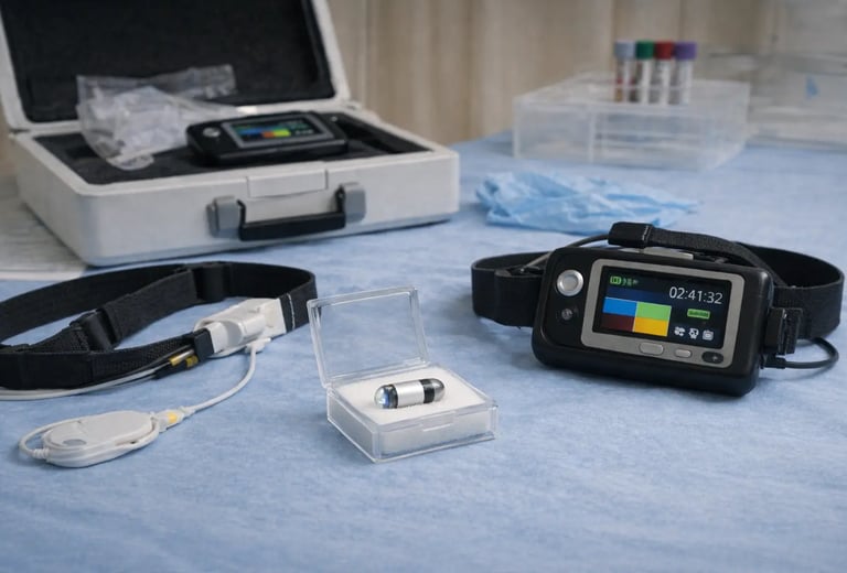
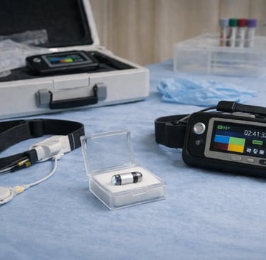
You arrive at your physician's office or endoscopy center in the morning. A technician attaches sensor patches or a sensor belt to your abdomen. These sensors receive the wireless signal from the capsule and feed data to the recording device.
The recorder - about smartphone-sized - clips to your belt or hangs from a shoulder strap. You'll wear it throughout the study.
Swallowing the Capsule
With a glass of water, you swallow the capsule like any other pill. Most people have no trouble. If swallowing is problematic, the capsule can sometimes be placed endoscopically into the stomach or small bowel.
Once swallowed, the capsule activates automatically. It starts capturing images as it travels through your esophagus.
During the Recording Period
Here's where capsule endoscopy differs dramatically from traditional procedures. After swallowing the capsule, you can leave. Go about your day.
You'll get instructions about when to resume drinking clear liquids (usually 2 hours after swallowing) and when to eat a light meal (typically 4 hours after). Avoid strenuous physical activity and heavy lifting. But normal daily activities are fine.
The capsule keeps recording for 8 to 12 hours until the battery dies. During this time, the recorder stores all transmitted images. You don't need to do anything except wear the device and stay away from MRI machines.
Returning the Equipment
At the end of the recording period - usually the same day - you go back to drop off the recorder and sensor equipment. The capsule itself stays inside you. It'll pass naturally.
What Does the Capsule Actually Record?
As the capsule travels through your digestive tract, it photographs the mucosal lining in vivid detail. The camera captures color images with significant magnification. Enough to visualize individual villi in the small intestine.
Normal Findings
Healthy small bowel mucosa appears pink with a velvety texture from the villi. Images show normal folds, blood vessel patterns and smooth transitions between intestinal segments.
Abnormal Findings
The capsule can detect a wide range of problems:
Ulcers and erosions - breaks in the mucosal surface ranging from tiny aphthous lesions to larger ulcerations
Erythema and edema - redness and swelling indicating inflammation
Angiodysplasias - abnormal blood vessels that can bleed
Active bleeding - fresh blood or clots visible in the intestinal lumen
Tumors and masses - growths projecting from or distorting the intestinal wall
Polyps - protruding lesions that may be benign or precancerous
Strictures - narrowed segments that may slow or block capsule passage
Villous atrophy - flattening of normal mucosal texture seen in celiac disease
Parasites - occasionally spotted when present
The capsule also documents transit times through different GI segments. That information can be clinically useful.
After the Study Is Done
Once you've returned the recording equipment, the data gets processed and reviewed.
Image Analysis
Specialized software downloads the images from the recorder. It compiles them into a video the physician can watch at adjustable speeds. The software also flags frames where the capsule detected red color - potentially indicating blood - and marks them for closer review.
A trained gastroenterologist watches the entire video. They examine each segment of bowel for abnormalities. This review typically takes 30 to 60 minutes. Depends on findings and image quality.
Getting Your Results
Results aren't immediate. Your physician needs time to review thousands of images carefully. Expect to wait several days to a week. Some cases take longer if the images need consultation with other specialists.
Your physician will contact you to discuss results. Or they'll schedule a follow-up appointment to go over them in person.
Capsule Passage
The capsule should pass in your stool within 24 to 72 hours after swallowing. Most people never notice it. You don't need to fish it out. Just flush normally.
If two weeks pass and you haven't seen the capsule, contact your physician. An abdominal X-ray can confirm whether it's still inside. Retained capsules occasionally need endoscopic or surgical removal. But that's uncommon.
Why Choose Capsule Endoscopy?
The procedure has several advantages over traditional approaches.
Noninvasive and Comfortable
No tubes getting passed through your body. No sedation. No recovery period. You swallow a pill and go about your day. For patients who find traditional endoscopy stressful, capsule endoscopy offers a gentler option.
Sees the Entire Small Bowel
Standard upper endoscopy reaches the duodenum. Colonoscopy with ileal intubation examines the terminal ileum. Everything between - roughly 18 feet of small intestine - remained invisible before capsule technology. Now physicians can see it all.
High Diagnostic Yield
For appropriate indications, capsule endoscopy frequently finds abnormalities that other tests miss. Multiple studies show superior sensitivity compared to small bowel barium studies, CT enterography and push enteroscopy when it comes to detecting mucosal lesions.
Safe with Few Side Effects
Serious complications are rare. The main risk is capsule retention. That occurs in about 1-2% of cases. Higher in patients with known Crohn's disease or suspected strictures. Most retained capsules pass on their own with time or respond to medical therapy.
Limitations Worth Knowing
Capsule endoscopy isn't perfect. Several limitations affect its utility.
No Therapeutic Capability
The capsule can only look. It can't take biopsies. Can't remove polyps. Can't cauterize bleeding vessels. Can't perform any interventions. If abnormalities show up, follow-up procedures may be necessary to get tissue or provide treatment.
Passive Movement
Unlike traditional endoscopy where the physician controls the scope's position, the capsule moves passively with peristalsis. It can't be steered, repositioned or slowed down. If it passes quickly through an area, visualization may be incomplete.
Variable Image Quality
Intestinal contents, bubbles and bile can obscure the view. Poor preparation leads to suboptimal images. Battery life sometimes runs out before the capsule completes its small bowel journey.
Capsule Retention Risk
In patients with strictures or obstructions, the capsule may get stuck. That's why careful patient selection and sometimes patency capsule testing are essential before the procedure.
Not a Colonoscopy Replacement
While colon capsule endoscopy exists, it's not recommended as routine screening for colorectal cancer. Standard colonoscopy remains superior because it allows polyp removal during the same procedure.
Who Shouldn't Have Capsule Endoscopy?
Certain conditions make capsule endoscopy inadvisable:
Known or suspected GI obstruction or stricture (unless patency testing confirms safe passage)
Swallowing disorders preventing pill ingestion
Pacemakers or defibrillators (relative contraindication - discuss with your physician)
Pregnancy (limited safety data)
Patients unable to follow preparation instructions
Your physician will evaluate whether capsule endoscopy makes sense for your specific situation.
What Happens After Results Come Back?
Results guide what comes next. Normal findings may reassure you that no significant small bowel pathology exists. Abnormal findings trigger additional workup.
If the capsule identifies a lesion needing biopsy or treatment, device-assisted enteroscopy (using balloon-assisted or spiral enteroscopes) can often reach the area. Some findings may require surgery.
For patients with Crohn's disease, capsule findings inform treatment decisions. Evidence of active inflammation despite therapy might prompt medication adjustments. Findings of mucosal healing may confirm treatment success.
The Technology Inside
The engineering packed into these tiny devices is remarkable. Each capsule contains:
One or two CMOS image sensors (cameras)
White LED light sources for illumination
A radiofrequency transmitter
Batteries providing 8 to 12+ hours of operation
An antenna
The cameras capture images through a transparent optical dome. LEDs illuminate the surrounding mucosa. The transmitter sends image data wirelessly to external sensors. Those relay it to the recording device.
Modern capsules achieve resolution good enough to detect lesions just a few millimeters across. Some systems offer adaptive frame rates that increase image capture when the capsule detects rapid movement.
Final Thoughts
Capsule endoscopy has opened a window into the small intestine that simply didn't exist two decades ago. For patients with obscure GI bleeding, suspected Crohn's disease, unexplained anemia and other small bowel conditions, it provides diagnostic information that was previously unobtainable without surgery.
The procedure is safe, noninvasive and well-tolerated. You swallow a pill. Wear a recorder for several hours. Go about your day. No sedation. No recovery time. No hospital stay.
If your physician has recommended capsule endoscopy, the procedure offers an excellent opportunity to visualize areas of your digestive tract that other tests simply can't reach.
© 2025. All rights reserved.
About Us
Introduction
Development
Cooperation
Service
Main Products
Medical Grade Monitor
No 15, Jinyang road KunshanSuzhou, Jiangsu, China