📞Customer Service: +86 13248368268 📧servicecenter@suzhoufrank.com one year replacement and warranty!

What Is Endoscopy? Explained
Everything you need to know about the endoscopy procedure, including preparation, sedation, recovery, and how it helps diagnose digestive issues.
ENDOSCOPY
Dr Qi Rui
12/15/202514 min read
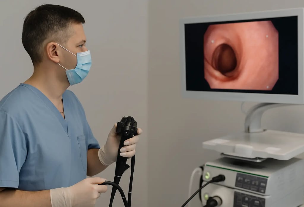
Endoscopy is a medical procedure that lets physicians examine the inside of your body using a flexible tube fitted with a miniature camera. This instrument - called an endoscope - gets inserted through a natural opening such as the mouth or rectum. It gives doctors a direct view of organs like the stomach, intestines and colon. No surgical incisions required.
The procedure serves two purposes. Physicians use it to pinpoint what's causing symptoms like abdominal pain, GI bleeding or dysphagia. And during the same session, they can often treat the problem directly - removing polyps, controlling hemorrhage or obtaining tissue for biopsy.
It's become one of the most frequently performed procedures in gastroenterology. There's a good reason for that. It offers a safe way to visualize the digestive tract and catches conditions early - everything from peptic ulcers to malignancies.
How Does an Endoscope Actually Work?
The endoscope itself is a thin flexible tube. Usually around 9 to 12 mm in diameter. At the distal tip sits a high-resolution camera alongside a light source. As the physician navigates the tube through your GI tract, the camera sends real-time images to a monitor.
This live visualization lets doctors inspect the mucosal lining of your esophagus, stomach and intestines with impressive clarity. Modern scopes produce HD images. They can pick up abnormalities just a few millimeters in size.
But here's what makes endoscopy particularly valuable. The tube contains working channels. Physicians pass specialized instruments through these channels to perform various interventions:
Obtaining tissue samples for histopathological analysis
Excising polyps or suspicious lesions
Injecting medications at the target site
Cauterizing bleeding vessels
Dilating strictures in the digestive tract
Deploying stents to maintain luminal patency
The Cleveland Clinic points out that this dual diagnostic and therapeutic capability is what makes endoscopy such a versatile tool in modern gastroenterology.
Different Types of Endoscopy Procedures
Something many patients don't realize - endoscopy isn't a single procedure. It's an entire family of procedures. Each one targets a specific anatomical region.
Upper Endoscopy (EGD)
Upper endoscopy - also called esophagogastroduodenoscopy - examines the esophagus, stomach and proximal small intestine (the duodenum). The physician passes the scope through your mouth and advances it down the throat. It's commonly indicated for investigating GERD, epigastric pain, nausea, vomiting and swallowing difficulties.
The procedure typically runs 15 to 30 minutes. Most patients receive sedation beforehand, so discomfort is minimal and recall is limited. If the physician spots anything concerning during the exam, they'll obtain a tissue sample right then for lab analysis. The full upper endoscopy procedure involves additional steps from prep through recovery. But that covers the core of it.
Colonoscopy
Colonoscopy provides visualization of the entire large intestine and rectum. The scope enters via the rectum and advances through the colon. It remains the gold standard for colorectal cancer screening. Current guidelines recommend all adults begin screening at age 45.
Patients often confuse these two procedures. The key distinction between endoscopy vs colonoscopy is simply which part of the GI tract gets examined. Upper endoscopy looks at everything proximal to the small bowel. Colonoscopy examines everything distal to it.
Sigmoidoscopy
This one focuses on just the lower colon - specifically the sigmoid colon. It covers less territory than a full colonoscopy, so it takes less time. Some physicians use it for targeted screening or to evaluate symptoms that seem localized to the lower GI tract.
Bronchoscopy
When physicians need to visualize the airways and lungs, they turn to bronchoscopy. The scope passes through the nose or mouth, down the throat and into the bronchial tree. It helps identify pulmonary infections, masses and other respiratory pathology.
Cystoscopy
Cystoscopy lets physicians examine the bladder and urethra. The scope enters through the urethral opening. Urologists rely on this procedure for investigating lower urinary tract symptoms, bladder abnormalities and urothelial malignancies.
ERCP
ERCP - endoscopic retrograde cholangiopancreatography - is more specialized. It combines endoscopy with fluoroscopic imaging to visualize the biliary tree, gallbladder and pancreatic duct. Physicians use it for both diagnosis and intervention. Retained common bile duct stones are a classic indication.
Capsule Endoscopy: A Different Approach
Standard endoscopy can't easily reach every part of the GI tract. The small intestine poses a particular challenge. It extends over 20 feet. That's difficult territory for a conventional scope.
Capsule endoscopy addresses this limitation.
Instead of a flexible tube, the patient swallows a pill-sized capsule housing one or two miniature cameras. Over 8 to 12 hours, it travels through the digestive system capturing upwards of 50,000 images. These transmit wirelessly to a recording device worn on the belt. Johns Hopkins Medicine notes that patients don't feel the capsule at all as it moves through their system.
The capsule eventually passes with a bowel movement. No retrieval necessary.
Understanding how capsule endoscopy works gives patients more confidence about this diagnostic modality. Many find it far more tolerable than conventional endoscopy - no sedation needed, minimal prep required.
Certain conditions capsule endoscopy can diagnose simply don't show up on other studies. The National Institutes of Health indicates it has superior diagnostic yield for small bowel pathology compared to barium studies, CT enterography or push enteroscopy. It excels at identifying obscure GI bleeding sources, small bowel neoplasms and ileal Crohn's disease.
Why Would a Patient Need an Endoscopy?
Physicians recommend endoscopy for three main reasons. To investigate symptoms. To screen for disease. Or to deliver treatment.
Investigating Symptoms
When a patient presents with persistent GI symptoms that don't resolve, an endoscopy test helps determine the underlying etiology. Common indications include:
Refractory heartburn unresponsive to PPI therapy
Odynophagia or dysphagia
Unexplained abdominal or epigastric pain
Persistent nausea and vomiting
Unintentional weight loss
Hematemesis or melena
Chronic diarrhea or constipation
Iron deficiency anemia of unclear origin
The range of what diseases endoscopy can detect is broad. GERD, peptic ulcer disease, celiac disease, inflammatory bowel disease, Barrett's esophagus, H. pylori infection - the list goes on. When patients ask what an endoscopy will show, the honest answer is it depends on their clinical picture. But the procedure delivers direct visual evidence that imaging studies and lab work simply can't provide.
Cancer Screening and Detection
Endoscopy serves a critical function in oncologic screening and early detection. Colonoscopy has dramatically reduced colorectal cancer mortality by enabling physicians to identify and resect premalignant polyps before they undergo malignant transformation.
Upper endoscopy aids in detecting esophageal and gastric malignancies. This proves especially important for patients with established risk factors - chronic reflux, Barrett's metaplasia or family history.
Patients frequently ask whether endoscopy can detect cancer. It can. And not just detect it. The procedure allows immediate tissue sampling from suspicious lesions for histopathologic confirmation. The American Cancer Society considers this combination of direct visualization plus biopsy capability one of the most reliable approaches for diagnosing GI malignancies.
Therapeutic Intervention
Endoscopy isn't purely diagnostic. Physicians can manage numerous conditions during the same session. Polypectomy. Esophageal dilation. Hemostasis of bleeding lesions. Stent placement for malignant obstruction. Foreign body retrieval.
This therapeutic capability spares patients additional procedures. And it often provides immediate symptom relief.
Preparing for Your Endoscopy
Adequate preparation ensures optimal visualization and reduces complication risk. Your care team will provide specific instructions based on which procedure you're undergoing.
For upper endoscopy, the main requirement is gastric emptying. You'll typically fast for 8 to 12 hours before the procedure. Clear liquids are usually permitted until 2 to 4 hours prior.
Colonoscopy demands more extensive prep. The colon must be thoroughly cleansed for the camera to adequately visualize the mucosa. This means a clear liquid diet the day before plus a bowel preparation solution to evacuate intestinal contents. Mayo Clinic stresses that prep quality directly affects examination adequacy. Poor prep means potentially missed pathology.
A thorough endoscopy preparation checklist generally includes:
Preparation Step Timing Review all current medications with physician 1 week prior Hold anticoagulants if directed 5 to 7 days prior Arrange post-procedure transportation Before procedure day Begin clear liquid diet Day before (colonoscopy) Complete bowel prep solution Evening before (colonoscopy) NPO for solid food 8 to 12 hours prior NPO for all liquids 2 to 4 hours prior
Disclose all medications to your physician. This includes OTC drugs and supplements. Anticoagulants may require temporary discontinuation. Diabetic patients often need medication adjustments given the fasting requirements.
You won't be fit to drive following the procedure. Sedation impairs judgment and reflexes for several hours. Arrange transportation in advance. Most facilities won't proceed without confirmed arrangements.
What Happens During the Procedure
Knowing the endoscopy procedure step by step helps alleviate pre-procedure anxiety.
Prior to the Examination
Plan to arrive 30 to 60 minutes before your scheduled time. Staff will confirm your identity, review your history and verify you've completed all prep instructions. You'll change into a gown. An IV line goes into your arm for fluid and medication administration.
Sedation
Most endoscopies employ moderate (conscious) sedation. You'll feel relaxed and drowsy. Many patients doze off and have little to no recollection afterward. The medications work quickly. Effects dissipate within an hour or two post-procedure.
One common concern - whether endoscopy is painful. With proper sedation, most patients report minimal discomfort. Some pressure or mild cramping perhaps. But significant pain is unusual. The care team monitors you continuously and can titrate sedation as needed.
The Examination Itself
Once adequately sedated, the physician advances the endoscope. For upper endoscopy, a bite block protects your dentition as the scope passes through the oropharynx. You maintain normal respirations throughout.
For colonoscopy, you're positioned on your side. The scope enters via the rectum. The physician may insufflate the colon with air or CO2 to improve mucosal visualization.
The examination itself proceeds fairly quickly. How long an endoscopy takes varies by procedure type and whether intervention is required. A straightforward EGD might take 10 to 15 minutes. Colonoscopy typically runs 20 to 40 minutes. Complex procedures like ERCP can exceed an hour.
The physician carefully inspects the tissue, documents abnormalities photographically and performs biopsies or therapeutic maneuvers as clinically indicated.
Recovery
Following scope withdrawal, you'll rest in a recovery area as sedation effects subside. This generally takes 30 to 60 minutes. Nursing staff monitor your vitals and confirm stability prior to discharge.
Mild endoscopy side effects may occur - bloating, flatulence, pharyngeal discomfort (after upper endoscopy). These typically resolve within 24 to 48 hours. WebMD notes that serious complications remain rare when trained specialists perform the procedure.
Your physician may share preliminary findings before discharge. Or they'll schedule follow-up once histopathology results return.
Understanding Your Endoscopy Results
So the procedure's done. Now what?
Your physician may share some preliminary observations before you leave the facility. They can tell you if the mucosa looked healthy, if they spotted any lesions or if they removed any polyps. But that's not the complete picture.
If biopsies were taken, the tissue samples go to a pathology lab. A pathologist examines them under a microscope looking for cellular abnormalities - inflammation, dysplasia, malignancy. This histopathologic analysis typically takes 3 to 7 business days. Sometimes longer for specialized staining or consultations.
Once results return, your physician will contact you. Or you'll have a scheduled follow-up appointment to discuss findings in detail. Don't panic if you don't hear back immediately. No news usually means the lab is still processing.
What might the results reveal? A few possibilities:
Normal findings. The examination showed healthy tissue throughout. No inflammation, no lesions, no concerning features. This is obviously the outcome everyone hopes for.
Inflammation. The mucosa appeared red, swollen or irritated. This could indicate gastritis, esophagitis, colitis or other inflammatory conditions. Treatment depends on the underlying cause.
Ulceration. Open sores in the GI lining. Peptic ulcers in the stomach or duodenum. Esophageal ulcers from severe reflux. These typically require acid suppression therapy and sometimes antibiotics if H. pylori is involved.
Polyps. Abnormal tissue growths projecting from the mucosal surface. Most colorectal polyps are benign. But certain types (adenomas) carry malignant potential. That's precisely why colonoscopy screening matters - catch them early, remove them, prevent cancer.
Masses or tumors. Suspicious growths requiring further evaluation. Biopsy results determine whether they're benign or malignant. This finding triggers additional workup - staging imaging, oncology referral, treatment planning.
Strictures. Narrowed segments of the GI tract. Could result from scarring, inflammation or malignancy. May require dilation or other intervention.
The key point here - endoscopy provides direct visualization that other tests can't match. Your physician actually sees what's happening inside. Combined with tissue sampling, it delivers diagnostic certainty.
Is Endoscopy Safe? Understanding the Risks
Patients understandably worry about complications. It's a medical procedure after all. Instruments going inside your body. Sedation involved.
Here's the reality. Endoscopy has an excellent safety profile. Millions of these procedures get performed annually with very low complication rates. But no medical procedure is completely without risk.
When patients ask is endoscopy dangerous, the honest answer is that serious complications are uncommon. But they can occur. Being informed helps you weigh benefits against risks.
Potential complications include:
Bleeding. This happens most often when biopsies are taken or polyps removed. Minor bleeding usually stops on its own. Significant hemorrhage requiring intervention is rare - occurring in roughly 1 in 1,000 procedures involving polypectomy.
Perforation. A tear in the GI tract wall. This is the complication physicians worry about most. It can require surgical repair. Fortunately it's rare. Upper endoscopy carries a perforation risk around 1 in 10,000. Colonoscopy slightly higher at 1 in 1,000 to 1 in 2,500.
Infection. Uncommon but possible. Endoscopes undergo rigorous disinfection protocols between patients. Still, infections can occasionally occur. Risk increases in certain procedures like ERCP.
Adverse reaction to sedation. Some patients experience respiratory depression, aspiration or allergic reactions to sedative medications. The anesthesia team monitors you closely to catch and manage these issues immediately.
Missed lesions. Not a complication per se. But worth mentioning. No diagnostic test is 100% sensitive. Small or flat lesions can occasionally escape detection. That's why surveillance intervals exist for high-risk patients.
Who faces higher risk?
Certain patient populations carry elevated procedural risk:
Elderly patients
Those with multiple comorbidities
Patients on anticoagulation therapy
Individuals with prior abdominal surgeries
Those with anatomical abnormalities
Patients requiring therapeutic intervention during the procedure
Your physician weighs these factors when recommending endoscopy. The benefits typically outweigh the risks. But it's always an individualized decision.
Recovery: What to Expect Afterward
Most patients tolerate endoscopy extremely well. Recovery is usually straightforward.
Immediately post-procedure, you'll spend 30 to 60 minutes in a recovery area. The sedation needs time to wear off. Nurses monitor your vital signs - blood pressure, heart rate, oxygen saturation. They'll make sure you're stable and alert before discharge.
What to eat after endoscopy depends on which procedure you had. Following upper endoscopy, your throat may feel slightly sore or scratchy. Start with soft foods and cool liquids. Avoid anything too hot or irritating for the first 24 hours. Most patients resume normal eating by the next day.
After colonoscopy, you might experience bloating and gas from the air used to inflate your colon. This passes. Literally. Walking around helps move things along. Start with light meals and advance as tolerated.
Common post-procedure symptoms:
Mild sore throat (upper endoscopy)
Bloating or cramping (colonoscopy)
Grogginess from sedation
Slight nausea
Fatigue
These typically resolve within 24 to 48 hours. Nothing to worry about.
Warning signs that require attention:
Contact your physician immediately if you experience:
Severe abdominal pain that worsens
Fever or chills
Persistent vomiting
Blood in your stool (more than slight streaking)
Vomiting blood or material that looks like coffee grounds
Difficulty swallowing that doesn't improve
Chest pain
These could indicate a complication requiring evaluation. Don't wait it out.
Activity restrictions:
The sedation impairs your reflexes and judgment for longer than you might realize. For the rest of procedure day:
Don't drive
Don't operate machinery
Don't make important decisions or sign legal documents
Don't consume alcohol
Most patients return to normal activities the following day. Some take an extra day off work just to be safe. Listen to your body.
Endoscopy Technology: How It Continues to Evolve
The field hasn't stood still. Endoscopy tech has advanced remarkably over the past two decades. And it keeps getting better.
High-definition imaging. Today's endoscopes capture images in stunning detail. HD and even 4K resolution is now standard in many facilities. This allows detection of subtle mucosal changes that older scopes would miss.
Narrow-band imaging (NBI). This technology uses specific wavelengths of light to enhance visualization of blood vessels and surface patterns. It helps distinguish between benign and potentially malignant tissue without requiring biopsy of every suspicious area.
Chromoendoscopy. Physicians spray dye onto the mucosal surface to highlight abnormalities. The dye pools in irregular areas, making lesions more visible. Particularly useful for detecting flat polyps and early cancers.
Endoscopic ultrasound (EUS). Combines endoscopy with ultrasound imaging. The scope has an ultrasound transducer at its tip. This allows visualization of structures beyond the GI wall - lymph nodes, pancreas, bile ducts. Essential for staging GI malignancies.
Confocal laser endomicroscopy. This is essentially microscopy in real-time during endoscopy. It provides cellular-level imaging of tissue. Physicians can essentially perform an "optical biopsy" during the procedure.
Artificial intelligence. AI-assisted detection systems are now entering clinical practice. These algorithms analyze endoscopic images in real-time and flag potentially abnormal areas. Studies show they can improve polyp detection rates.
Robotic endoscopy. Still largely experimental. But robotic platforms are being developed that could allow more precise navigation and intervention. The future looks interesting.
The bottom line - endoscopy equipment continues to improve. Better optics. Better detection. Better outcomes for patients.
Advanced Endoscopy: When Standard Procedures Aren't Enough
Sometimes conventional endoscopy doesn't provide adequate access or therapeutic capability. That's where advanced endoscopy comes in.
Advanced endoscopy meaning refers to specialized techniques requiring additional fellowship training beyond standard gastroenterology. These procedures tackle more complex clinical scenarios.
Double-balloon enteroscopy. Remember how we said the small intestine is hard to reach? This technique uses an endoscope with two inflatable balloons that allow the physician to "inch" through the small bowel. It can reach areas capsule endoscopy identifies as abnormal but can't treat.
Endoscopic mucosal resection (EMR). A technique for removing larger or flatter lesions that can't be snared with standard polypectomy. The physician injects fluid beneath the lesion to lift it, then resects it.
Endoscopic submucosal dissection (ESD). Takes EMR further. Allows en bloc removal of even larger lesions. Originated in Japan for early gastric cancer treatment. Now used more widely.
POEM procedure. Peroral endoscopic myotomy. Used to treat achalasia - a swallowing disorder where the lower esophageal sphincter doesn't relax properly. The physician tunnels under the esophageal lining and cuts the muscle fibers. No external incisions.
Endoscopic necrosectomy. For patients with infected pancreatic necrosis. The physician creates an opening between the stomach and the necrotic collection, then removes dead tissue endoscopically. Avoids major surgery.
These techniques represent the cutting edge. They require specialized training and equipment. But they offer treatment options that didn't exist a generation ago.
Gastroscopy vs Endoscopy: Clearing Up the Confusion
Patients sometimes get confused by terminology. They hear "gastroscopy" and "endoscopy" and wonder if they're different procedures.
The distinction with gastroscopy vs endoscopy is mostly semantic. Gastroscopy is simply a specific type of endoscopy. It examines the stomach (gastro = stomach). The term is used interchangeably with upper endoscopy or EGD in many countries.
Think of it this way. Endoscopy is the umbrella term. Gastroscopy, colonoscopy, bronchoscopy, cystoscopy - these are all specific types of endoscopy targeting different organs.
In practice, when a physician says "endoscopy" without further specification, they usually mean upper endoscopy/gastroscopy. Context matters.
Cost Considerations
Let's address something patients often wonder about but hesitate to ask. Endoscopy cost varies considerably depending on several factors.
What affects the price?
Geographic location. Costs differ significantly between regions and countries. Urban centers tend to be pricier than rural areas.
Facility type. Hospital-based endoscopy typically costs more than ambulatory surgery centers or office-based procedures. Overhead differences explain much of this gap.
Type of procedure. A straightforward diagnostic EGD costs less than a complex therapeutic ERCP. More time, more equipment, more specialized expertise - higher cost.
Anesthesia. Monitored anesthesia care (MAC) with an anesthesiologist present costs more than moderate sedation administered by the endoscopist.
Pathology. If biopsies are taken, lab fees add to the total. Multiple biopsies mean multiple charges.
Insurance coverage. This is the big variable. With insurance, your out-of-pocket might be minimal. Without insurance, you're looking at the full facility fee, professional fee, anesthesia fee and pathology fee. It adds up.
Ballpark figures:
In the United States, an upper endoscopy might range from $1,000 to $3,000 or more depending on the above factors. Colonoscopy often runs $1,500 to $4,500. These are rough estimates. Actual costs vary widely.
The important thing - don't let cost concerns prevent you from getting a necessary procedure. Talk to your physician and the facility's billing department. Payment plans often exist. Some facilities offer cash-pay discounts.
Frequently Asked Questions
Can I eat before endoscopy?
No. Your stomach needs to be empty. Typically you'll fast for 8 to 12 hours before upper endoscopy. Colonoscopy requires clear liquids the day before plus complete bowel prep.
Will I be asleep during the procedure?
Most patients receive moderate sedation. You'll be drowsy and relaxed. Many patients doze off and don't remember the procedure. General anesthesia (completely unconscious) is used only in specific circumstances.
How soon can I return to work?
Most patients take procedure day off and return to work the following day. If you have a physically demanding job, you might want an extra day. Listen to how you feel.
How often should I get a colonoscopy?
For average-risk individuals, every 10 years starting at age 45. If polyps are found or you have risk factors, more frequent surveillance applies. Your physician will recommend an appropriate interval.
Can endoscopy miss cancer?
No test is perfect. Small or flat lesions can occasionally escape detection. However, endoscopy remains the most accurate method for visualizing and sampling GI tract abnormalities. The miss rate for significant lesions is low in experienced hands.
Is the bowel prep really that bad?
It's not pleasant. You'll spend significant time in the bathroom. The taste of prep solutions has improved over the years. Most patients manage fine. The key is following instructions carefully and staying hydrated.
The Role of Quality Equipment in Endoscopy
One aspect patients rarely consider - the equipment matters. A lot.
High-quality endoscopes produce clearer images. Better optics mean better detection of subtle abnormalities. Reliable equipment means fewer technical issues during procedures. This translates directly to patient outcomes.
Healthcare facilities invest significantly in endoscopy equipment. The cameras, light sources, processors and accessories all contribute to procedural quality. Modern disposable components have also improved safety and reduced cross-contamination risks.
For healthcare facilities looking to upgrade their endoscopy capabilities, camera technology represents a critical component. Superior imaging helps clinicians detect lesions earlier and perform interventions more precisely.
If you're a healthcare provider exploring equipment options, Suzhou Frank Medical offers high-quality disposable endoscope cameras designed for reliable performance and patient safety. These single-use camera systems eliminate cross-contamination concerns while delivering the image quality clinicians need for accurate diagnosis and treatment.
© 2025. All rights reserved.
About Us
Introduction
Development
Cooperation
Service
Main Products
Medical Grade Monitor
No 15, Jinyang road KunshanSuzhou, Jiangsu, China