📞Customer Service: +86 13248368268 📧servicecenter@suzhoufrank.com one year replacement and warranty!

What Will an Endoscopy Show? Results Explained
What will an endoscopy show? Learn what the procedure reveals about ulcers , inflammation , Barrett's esophagus , polyps and other digestive tract conditions.
ENDOSCOPY
Dr Qi Rui
12/21/20255 min read
What will an endoscopy show? It's one of the most common questions people ask after their doctor orders the procedure. Healthcare professionals hear this question constantly , and the answer shapes how patients understand their diagnosis and treatment path.
The short answer is that endoscopy shows the actual lining of the digestive tract in real time. Unlike X-rays or CT scans that produce indirect images , endoscopy provides direct visualization. Physicians see the tissue itself , not shadows or outlines. They can identify inflammation , ulcers , bleeding sources , structural problems and suspicious growths. They can also take tissue samples for laboratory analysis when something needs closer examination.
What an endoscopy can show depends partly on why it was ordered. Someone with chronic heartburn will have different potential findings than someone with difficulty swallowing or unexplained weight loss. But across all cases , endoscopy answers questions that other tests simply can't address with the same clarity.
How Endoscopy Captures What's Inside
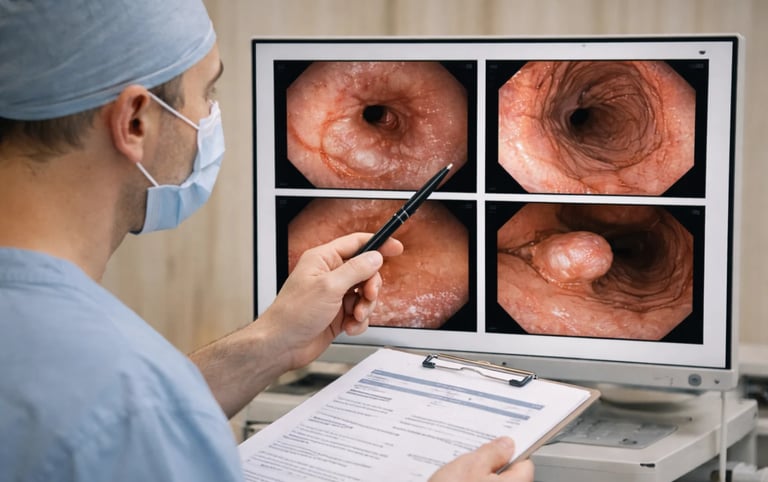
Understanding what endoscopy involves helps explain why it reveals so much. A thin flexible tube with a camera at the tip passes through the mouth and into the digestive tract. The camera transmits live images to a monitor , allowing the physician to examine the esophagus , stomach and first part of the small intestine (duodenum) in detail.
The quality of what an endoscopy shows depends significantly on imaging technology. Modern high-definition systems capture subtle details that older equipment might miss. Facilities using ultra HD 4K medical endoscope cameras can detect fine vascular patterns , slight color variations and early-stage abnormalities with greater precision. For both patients and clinicians , better imaging means more accurate diagnosis.
During the examination , the physician evaluates tissue color , texture , structural integrity and any focal abnormalities. Normal mucosa appears smooth , pink and uniform. Anything deviating from this baseline warrants closer inspection and often tissue sampling.
What Can an Endoscopy Show in the Esophagus?
The esophagus is examined first as the scope passes from the throat toward the stomach. Several conditions show characteristic appearances on endoscopy.
Acid reflux damage (esophagitis) appears as redness , erosions or ulcerations in the lower esophagus. The severity ranges from minimal irritation to extensive damage. What endoscopy shows in reflux patients helps determine whether lifestyle changes alone will work or whether more aggressive treatment is needed. The American College of Gastroenterology emphasizes that endoscopic findings directly guide treatment intensity for reflux disease.
Barrett's esophagus shows as salmon-colored tissue extending upward from where the esophagus meets the stomach. This condition develops in some people with long-standing reflux and carries increased cancer risk. Finding Barrett's on endoscopy triggers a surveillance program with regular follow-up examinations.
Strictures appear as narrowed areas that may cause swallowing difficulty. Endoscopy shows whether the narrowing looks benign (smooth and symmetric) or suspicious for malignancy (irregular with mass-like features). Tissue sampling helps confirm the diagnosis.
Esophageal varices are enlarged veins that indicate liver disease with portal hypertension. What an endoscopy shows regarding varice size and appearance directly determines bleeding risk and guides preventive treatment decisions.
Cancer may appear as masses , ulcerated areas or irregular strictures. Early esophageal cancer can look subtle , sometimes presenting as slight discoloration or minimal irregularity. This is where image quality becomes critical for detection.
What Does an Endoscopy Show in the Stomach?
The stomach offers a larger surface area to examine. Physicians follow systematic patterns to ensure complete visualization of all regions.
Gastritis (stomach inflammation) shows as redness , swelling or erosions. What endoscopy shows can suggest the underlying cause , though biopsy often provides confirmation. Helicobacter pylori infection , NSAID use , alcohol and autoimmune conditions all produce recognizable patterns.
Ulcers appear as craters or breaks in the stomach lining. Endoscopy shows ulcer size , depth , location and any signs of recent or active bleeding. These visual characteristics determine urgency of treatment. The Mayo Clinic notes that endoscopy remains the gold standard for ulcer diagnosis and assessment.
Polyps are growths projecting from the stomach wall. What an endoscopy shows about polyp appearance helps predict whether they're benign or potentially precancerous. Size , shape and surface characteristics all matter. Most gastric polyps are harmless , but certain types require removal and follow-up.
Stomach cancer can present in various ways: as masses , ulcers that don't look typical , or thickened rigid walls. Early gastric cancer may appear subtle , making thorough examination and high-quality imaging essential.
Bleeding sources become visible during endoscopy when patients present with signs of GI hemorrhage. Endoscopy shows exactly where bleeding originates , whether it's actively continuing and what intervention might stop it.
What Will an Endoscopy Show in the Duodenum?
The duodenum (first part of the small intestine) is examined last during standard upper endoscopy.
Duodenal ulcers are common findings , strongly associated with H. pylori infection or NSAID use. Unlike stomach ulcers , duodenal ulcers rarely indicate cancer.
Celiac disease produces characteristic changes visible on endoscopy: scalloped folds , reduced fold number and a mosaic-like mucosal pattern. However , what an endoscopy shows visually isn't enough for diagnosis. Tissue biopsies confirming villous atrophy remain essential , even when the appearance seems classic.
Duodenitis (inflammation) appears as redness or erosions and has various causes including infection , medication effects and inflammatory conditions.
Beyond What the Eye Can See: Tissue Sampling
What endoscopy shows visually represents only part of the diagnostic picture. Tissue sampling extends capability significantly.
Biopsy involves taking small tissue samples for microscopic examination. This confirms what visual findings suggest , identifies infections , grades the severity of conditions like Barrett's esophagus and distinguishes benign from malignant processes. Standard practice includes sampling any suspicious areas and sometimes normal-appearing tissue when specific conditions are suspected.
For patients , understanding that biopsy is routine helps reduce anxiety. Taking samples doesn't mean cancer is suspected. It's often just thorough medicine.
For clinicians , biopsy protocols vary by indication. Barrett's surveillance follows specific mapping patterns. H. pylori testing requires samples from particular gastric locations. Celiac evaluation needs adequate duodenal specimens. Proper technique and specimen handling affect diagnostic accuracy.
What Endoscopy Results Mean
After the procedure , results come in stages. The physician can often share preliminary visual findings immediately: what the endoscopy showed in terms of overall appearance , any obvious abnormalities and whether everything looked normal.
Biopsy results take longer , typically several days to a week. These provide definitive answers about tissue characteristics , infection presence and cellular abnormalities.
Normal findings are genuinely reassuring. When endoscopy shows healthy-appearing mucosa throughout , it effectively rules out many concerning conditions. This negative result has real value.
Abnormal findings range from minor to significant. Mild gastritis might need only lifestyle modification. Barrett's esophagus requires surveillance planning. Suspicious masses demand prompt follow-up and often additional testing.
For healthcare teams , structured documentation of what endoscopy shows facilitates care coordination. Photographic records , standardized terminology and clear recommendations support continuity across providers.
Factors Affecting What Endoscopy Can Show
Several factors influence diagnostic yield.
Preparation quality matters. Residual food in the stomach obscures visualization. Following fasting instructions ensures the clearest possible view.
Equipment capability affects detection. Modern HD and 4K systems reveal subtle findings that standard-definition equipment might miss. In some settings , portable endoscopy systems bring high-quality imaging to locations outside traditional endoscopy suites , expanding access without sacrificing diagnostic accuracy.
Examiner experience influences outcomes. Skilled endoscopists detect subtle abnormalities more reliably. Adequate examination time correlates with improved findings , particularly for early-stage conditions.
Patient factors including anatomy , tolerance and sedation response can affect how thoroughly the examination proceeds.
When Endoscopy Shows Nothing Abnormal
A normal endoscopy result is valuable information. It effectively excludes structural problems , significant inflammation , ulcers and visible tumors. For patients with concerning symptoms , knowing what endoscopy didn't show can be as important as positive findings.
However , normal visual appearance doesn't rule out all conditions. Functional disorders like gastroparesis or functional dyspepsia may show no visible abnormalities. Motility problems require different testing. Some early-stage conditions may appear normal on standard examination.
When endoscopy shows normal findings but symptoms persist , further evaluation may be needed. This doesn't mean the procedure failed. It successfully narrowed the diagnostic possibilities.
Making Sense of Your Results
For patients , discussing endoscopy findings with the physician provides necessary context. What the endoscopy showed matters , but so does what it means for the individual situation. Treatment recommendations flow from the specific findings combined with symptoms , medical history and overall clinical picture.
For healthcare professionals , endoscopy remains irreplaceable for direct mucosal visualization. What it shows guides decisions across the full spectrum of upper GI conditions. Understanding both capabilities and limitations helps in ordering appropriate tests , setting realistic expectations and interpreting results accurately.
The procedure answers the fundamental question it's designed to address: what does the digestive tract lining actually look like? That direct answer , whether reassuring or revealing problems , provides the foundation for moving forward with appropriate care.
© 2025. All rights reserved.
About Us
Introduction
Development
Cooperation
Service
Main Products
Medical Grade Monitor
No 15, Jinyang road KunshanSuzhou, Jiangsu, China