📞Customer Service: +86 13248368268 📧servicecenter@suzhoufrank.com one year replacement and warranty!

The Importance of Surgical Cameras in Modern Medicine
Surgical camera technology transformed modern surgery. Understand how medical cameras work , why resolution matters and what to consider when selecting systems.
ENDOSCOPY
Dr Qi Rui
1/14/20266 min read
Surgery has transformed dramatically over the past few decades. What once required large incisions and direct visualization now happens through tiny openings guided by sophisticated imaging technology. At the heart of this transformation sits the surgical camera , an instrument so fundamental to contemporary practice that modern surgery would be unrecognizable without it.
Understanding how surgical cameras and medical cameras function , why they matter and what distinguishes quality systems from adequate ones helps clinicians , facility administrators and patients appreciate the technology enabling today's minimally invasive procedures.
From Direct Vision to Digital Visualization
Traditional open surgery relied on the surgeon's direct line of sight. Large incisions provided access and visibility simultaneously. The surgeon's eyes were the primary imaging system , limited only by lighting conditions and physical access to the operative field.
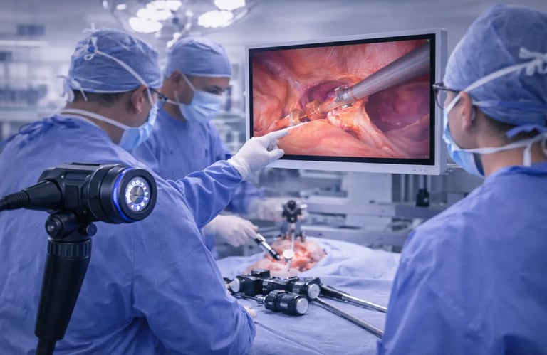
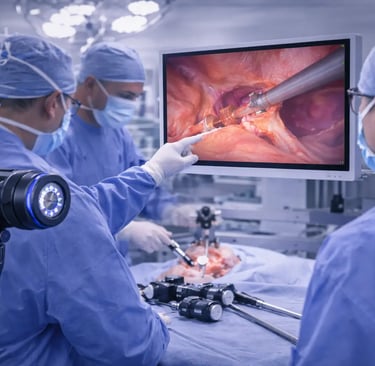
Minimally invasive surgery changed this equation entirely. When procedures happen through small ports and narrow channels , direct visualization becomes impossible. The surgical camera becomes the surgeon's eyes , capturing images from inside the body and displaying them on monitors where the entire team can see.
This shift represents more than technical evolution. It fundamentally altered what surgery can accomplish. Procedures that once required week-long hospital stays now happen as outpatient cases. Recovery measured in months shortened to days. Complications from large incisions , including infection , hernia and prolonged pain , diminished dramatically.
The American College of Surgeons documents how minimally invasive techniques have expanded across virtually every surgical specialty. This expansion depends entirely on medical camera technology capable of revealing what surgeons need to see through increasingly small access points.
How Surgical Cameras Work
A surgical camera captures images through a lens system , converts light into electronic signals and transmits those signals to processing units that render viewable images on displays. The basic principle mirrors consumer cameras , but the requirements differ substantially.
Medical cameras must function in challenging environments. Body cavities are dark , requiring powerful illumination delivered through fiber optics or LED systems. Tissues are wet and reflective , demanding optical systems that handle glare and maintain color accuracy. Working distances are short and variable , requiring lenses that maintain focus across a range of depths.
The camera head attaches to an endoscope or laparoscope that enters the body. In flexible endoscopy , the camera may sit at the scope's tip , capturing images as it navigates curved pathways through the digestive tract. For a foundational understanding of how this works in gastrointestinal applications , reviewing what endoscopy involves provides helpful context.
In rigid laparoscopy , the camera typically attaches to the external end of a straight telescope inserted through a port. The optical system transmits the image through the scope to the camera head outside the body.
Signal processing converts raw camera data into viewable images. This processing affects color reproduction , contrast , noise reduction and image enhancement. Quality processing reveals tissue characteristics that inferior systems obscure.
Resolution and Image Quality
Resolution determines how much detail a surgical camera can capture and display. Higher resolution means more pixels , which translates to finer anatomical detail visible on the monitor.
Standard definition cameras dominated early minimally invasive surgery. While revolutionary at the time , SD resolution now appears inadequate for procedures requiring precise tissue discrimination. High definition brought substantial improvement , revealing structures invisible at lower resolutions.
4K resolution represents the current premium standard. With four times the pixel count of HD , 4K surgical cameras display tissue planes , vascular structures and pathological changes with remarkable clarity. Surgeons report improved ability to identify critical anatomy and distinguish normal from abnormal tissue.
The Society of American Gastrointestinal and Endoscopic Surgeons recognizes visualization quality as fundamental to safe minimally invasive surgery. Their guidelines address how imaging technology affects procedural safety and outcomes.
Resolution alone doesn't determine image quality. Color accuracy ensures tissues appear as they actually are , enabling discrimination between tissue types based on color differences. Dynamic range affects how well the system handles brightness variation , from dark recesses to reflective surfaces illuminated directly. Frame rate determines motion smoothness , critical when instruments and tissues move during active surgery.
Illumination: The Partner to Every Medical Camera
A surgical camera can only capture what light reveals. Illumination quality directly affects image quality regardless of camera resolution.
Early endoscopic systems used external light sources connected by fiber optic cables. Light traveled from the source through flexible bundles to the scope tip , illuminating the field for camera capture. This approach remains common , with modern LED light sources offering consistent color temperature , long life and reliable output.
LED technology has largely replaced older halogen and xenon sources for routine applications. LEDs offer instant-on capability , minimal heat generation , stable color output and energy efficiency. For high-intensity applications , xenon sources still find use , but LED systems continue improving and expanding into demanding applications.
Illumination must match the surgical camera's capabilities. A 4K camera paired with inadequate lighting won't deliver 4K quality images. The entire imaging chain , from light source through camera to display , must maintain quality for the system to achieve its potential.
Types of Surgical and Medical Cameras
Different surgical applications require different camera configurations. Understanding the options helps match equipment to clinical needs.
Endoscope cameras integrate with flexible scopes for gastrointestinal , pulmonary and other applications where navigating curved anatomy is required. These medical endoscope cameras must be compact enough to fit scope dimensions while delivering resolution adequate for diagnostic and therapeutic work. Modern systems achieve HD and increasingly 4K resolution in remarkably small form factors.
Laparoscopic cameras attach to rigid telescopes for abdominal and pelvic surgery. These systems can be larger since the camera head remains outside the body. This allows more sophisticated optics and imaging chips , often delivering the highest resolution available in surgical imaging.
Portable camera systems serve facilities needing flexibility. Portable endoscope camera systems combine camera , processor and display in mobile configurations that can move between procedure rooms or to bedside locations. While sometimes sacrificing features available in fixed installations , portable systems extend surgical camera capability to settings where permanent infrastructure isn't practical.
3D camera systems capture stereoscopic images providing depth perception. Traditional 2D displays show flat images , requiring surgeons to infer depth from indirect cues. 3D systems restore natural depth perception , potentially improving precision for complex tasks. The newest naked-eye 3D medical-grade monitors display stereoscopic images without requiring special glasses , eliminating a longstanding barrier to 3D adoption.
Robotic surgery cameras integrate with surgical robots , providing visualization for console-based surgeons controlling remote instruments. These systems typically offer 3D imaging and exceptional resolution given the precision robotic surgery demands.
Clinical Impact of Camera Quality
Does camera quality actually affect patient outcomes? Evidence suggests it does , though isolating the camera's contribution from other factors presents research challenges.
Surgeons consistently report that better visualization improves their ability to identify anatomy , preserve important structures and recognize pathology. When surgeons see more clearly , they can operate more precisely. Precision reduces complications.
The learning curve for new procedures shortens with better imaging. Trainees develop skills faster when they can see what their supervisors see clearly. Educational value extends beyond the trainee at the scope , with high-quality imaging allowing entire teams to observe and learn.
Diagnostic accuracy improves with enhanced visualization. Medical cameras used for endoscopic diagnosis reveal findings that lower-quality systems miss. Early cancer detection , identification of subtle lesions and characterization of mucosal abnormalities all benefit from superior imaging.
Documentation quality affects care beyond the immediate procedure. Recorded images and video support consultation , patient education and quality review. High-resolution recording preserves detail that standard-definition capture loses.
Selecting Surgical Camera Systems
Facilities evaluating surgical camera options should consider several factors beyond basic specifications.
Clinical applications determine required capabilities. A facility performing complex minimally invasive surgery benefits from premium 4K systems. A clinic doing basic diagnostic endoscopy may find HD adequate and more cost-effective. Matching investment to clinical need avoids both overspending and underequipping.
Integration requirements affect practical utility. Cameras must work with existing scopes , light sources , recording systems and displays. Compatibility verification prevents expensive mismatches. Facilities with diverse equipment may value camera systems offering broad compatibility.
Ergonomics and handling matter for daily use. Camera heads that are comfortable to hold , intuitive to operate and appropriately weighted for their application reduce fatigue and improve procedural efficiency.
Reliability and support affect long-term value. Surgical cameras experience demanding use in challenging environments. Quality construction , manufacturer support and service availability influence total cost of ownership beyond purchase price.
Upgrade pathways deserve consideration given technology's continued evolution. Systems designed for component upgrades may offer better long-term value than those requiring complete replacement as technology advances.
The Suzhou Frank Medical Advantage
At Suzhou Frank Medical , we provide comprehensive surgical camera and medical camera solutions designed for clinical excellence. Our product range spans from flexible endoscope cameras for diagnostic applications to complete visualization systems incorporating cameras , light sources and displays.
What sets us apart is our integration of quality components across the entire imaging chain. Our cameras paired with matched illumination and medical-grade displays deliver consistent results that mismatched components cannot achieve. We focus exclusively on medical-grade equipment , ensuring our systems meet the demands of clinical environments rather than adapting consumer technology for medical use.
Explore our complete product catalog to find options suitable for facilities ranging from clinics establishing first endoscopy capability to hospitals upgrading existing surgical suites with current technology.
Looking Forward
Surgical camera technology continues advancing. Resolution will increase further. Processing will become more sophisticated. Artificial intelligence integration will help identify structures , flag abnormalities and assist surgical decision-making.
What won't change is the fundamental importance of visualization to modern surgery. As procedures become more complex and access becomes more minimal , the surgical camera's role only grows. Investing in quality imaging technology today positions facilities for both current excellence and future advancement.
Conclusion
The surgical camera transformed what surgery can accomplish. From basic diagnostic visualization to complex therapeutic procedures , medical cameras enable interventions that direct vision could never support. Understanding how these systems work , what distinguishes quality from adequacy and how to select appropriate equipment helps facilities deliver the best possible care.
© 2025. All rights reserved.
About Us
Introduction
Development
Cooperation
Service
Main Products
Medical Grade Monitor
No 15, Jinyang road KunshanSuzhou, Jiangsu, China